For decades, a cancer diagnosis has often pushed patients toward a familiar set of choices. Surgeons could cut. Radiation teams could aim beams at diseased tissue. Chemotherapy could move through the body in search of malignant cells. Each path has saved lives, yet each can leave patients facing pain, fatigue, scarring or long recovery.
A newer approach now sits beside those older tools for certain liver tumors, and its method sounds almost impossible at first. It does not begin with an incision. It does not depend on heat. It does not send radiation through the body. Instead, doctors use focused sound waves from outside the skin to destroy tumor tissue inside the liver.
Federal regulators cleared the system for liver tumor destruction in the United States, giving hospitals a path to use the treatment in selected patients. Behind that clearance sits a story that joins engineering, cancer medicine, imaging and a strange physical effect inside tissue. Tiny bubbles appear, collapse and leave the tumor broken apart.
One point needs care. Histotripsy is not a broad cure for cancer. It is a medical treatment now used for certain liver tumors, with doctors still studying where else it may work.
What The FDA Clearance Means
Histotripsy reached public attention after the U.S. Food and Drug Administration allowed HistoSonics to market its Edison system for noninvasive destruction of liver tumors. HistoSonics grew out of work at the University of Michigan, where engineers and doctors helped move the idea from research labs toward human use.
FDA clearance does not mean every cancer patient can receive the treatment. Doctors must decide case by case, using scans, blood tests, tumor measurements and the patient’s medical history. Location matters because ultrasound must reach the target clearly. Size matters because larger tumors may require more planning. Liver function matters because any liver procedure requires reserve capacity in the organ.
For patients who qualify, a hospital can use a commercial sound-wave system to destroy targeted liver tissue without placing a probe or blade into the tumor. Primary liver tumors and tumors that spread to the liver from another cancer site may fall within the population doctors review.
How Sound Waves Can Break A Tumor Apart
As the bubbles grow and burst, they place mechanical stress on nearby tumor cells. Instead of burning tissue, freezing it or poisoning it with drugs, histotripsy tears the tumor structure apart. Source material describes the treated mass as broken down into debris that the immune system and body can clear over time.
Precision drives the method. Energy from the device converges at the planned target, while physicians use imaging to monitor tissue during treatment.
Why Doctors Can Watch The Treatment In Real Time
Imaging helped move histotripsy into clinical use. Doctors use ultrasound imaging during planning and treatment, similar in principle to the scans many people associate with pregnancy. Here, imaging helps locate the tumor, map its edges and observe the treatment zone as bubbles form inside the target.
Real-time viewing matters because cancer treatment can depend on millimeters. A liver contains blood vessels, bile ducts and nearby organs. A doctor needs to know where destructive energy will go before treatment begins and how tissue responds once treatment starts.
HistoSonics’ Edison platform uses a treatment head connected to a robotic system. A soft water-filled interface sits against the patient’s abdomen so the sound waves can travel from the machine into the body. Once imaging locates the tumor, the team programs the target area and delivers the acoustic pulses.
Professor Tze Min Wah, a senior consultant interventional radiologist involved in the UK trial, described the lock-on step in plain terms. “Once the tumour in the liver has been located, it becomes the target which the system locks on to.”
What A Patient May Experience During The Procedure
Patients usually receive general anesthesia. Stillness helps the team aim accurately, and anesthesia prevents movement from breathing discomfort or muscle tension. Doctors may ask patients to follow dietary instructions before the appointment.
During treatment, the patient lies beneath the machine while the sound-wave head rests over the abdomen. A care team reviews imaging, confirms the target and delivers energy. Source material gives two timing ranges. A full appointment can last one to three hours, while active treatment may take only minutes for some tumors and longer for others.
Recovery can look very different from open surgery. No incision means no surgical wound. No probe means no needle track through the abdomen. Some patients may go home the same day, while others may stay overnight if doctors want to monitor pain, bleeding risk or liver response.
How Histotripsy Differs From Other Treatments
Traditional liver tumor care includes surgery, radiation, chemotherapy, immunotherapy and ablation. Surgeons may remove part of the liver when a tumor can be cut out safely. Ablation can destroy tumor tissue with heat or another local method, often through a needle placed into the tumor.
Histotripsy differs because it destroys tissue from outside the body with mechanical sound-wave force. No incision separates skin and muscle. No heat cooks the tumor. No radiation passes through tissue along a beam path. No drug compatibility issue arises from the sound waves themselves.
Such differences do not make older treatments obsolete. Many patients will still need surgery, chemotherapy, immunotherapy, radiation or thermal ablation. Doctors may use histotripsy when tumor size, number, position and health factors make the treatment reasonable.
Who May Be A Candidate

Current clinical use centers on liver tumors. Sources describe primary liver cancer, such as hepatocellular carcinoma, and metastatic tumors that have spread to the liver from cancers such as colon cancer or neuroendocrine tumors. Some medical centers mention symptomatic benign liver growths as another possible category.
Eligibility depends on practical anatomy. A tumor must be visible enough on ultrasound for doctors to aim safely. Ribs, gas or depth can block the beam. Tumors near sensitive structures may need extra care or a different method. Some hospitals describe ideal cases as three or fewer tumors, smaller than three centimeters and in favorable locations.
Doctors do not rely on one scan alone. A care team may review CT or MRI results, ultrasound visibility, blood work, liver function, clotting risk and prior treatment history. A patient who has been told surgery carries high risk may still need that full review before histotripsy enters the plan.
Side Effects And Risks Still Matter
Noninvasive does not mean risk-free. Histotripsy destroys tissue, and any treatment that destroys tissue can bring pain, bleeding, infection or injury outside the planned zone. General anesthesia also carries its own concerns, especially for patients with heart, lung or other health issues.
Many patients may feel mild pain or soreness after treatment. Doctors may keep someone overnight if pain, medical history or tumor location raises concern. Research still needs to answer long-term questions about recurrence, survival and best treatment combinations.
Trial Results And The Immune System Question
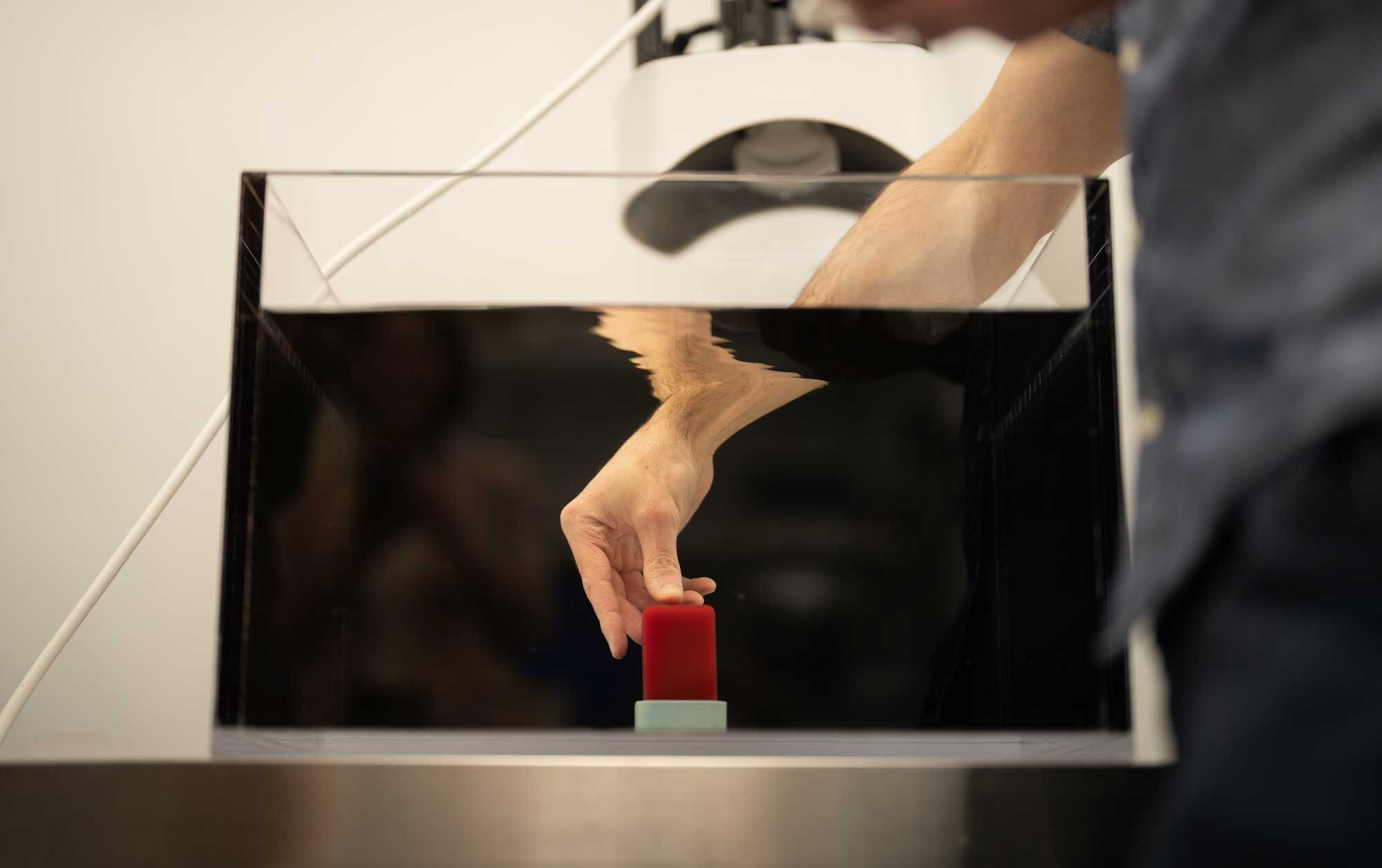
Human trials helped support early use of the Edison system for liver tumors. University of Michigan and other sites treated patients with primary and metastatic liver tumors, and the trial met stated safety and effectiveness goals in the source material. Early clinical use can show feasibility and safety signals, yet cancer care relies on longer follow-up.
Researchers also study what happens after sound waves rupture cancer cells. Preclinical rodent studies described in the source material suggest that, after partial tumor destruction, immune responses may help clear remaining disease in some animals. Scientists want to know if exposed tumor antigens can help immune cells recognize cancer more effectively, but that question still needs human data.
Professor Wah described what happens to treated tissue after the sound waves break it apart. “As the debris of the tumour has been liquified, it will be absorbed naturally by the body before passing out of it as waste,” she said.
What The Approval Really Signals
Histotripsy changes the menu of liver tumor treatment by giving doctors a way to destroy selected tumors without cutting into the body.
Medical progress often arrives with both hope and limits. For a suitable liver tumor, focused sound waves may mean less pain, faster recovery and no incision. For another patient, tumor position or disease spread may make surgery, ablation, radiation or medicine the better route.
FDA clearance brought the technology into real hospital use for liver tumors, but the next chapter will depend on careful patient selection, long-term data and honest conversations between doctors and patients. Sound waves can now destroy certain liver tumors. How far that idea can go remains a question medicine is still testing.
Featured Image Source: Erica Bass, Rogel Cancer Center, Michigan Medicine
https://medschool.umich.edu/health-lab/histotripsy-liver-tumor-trial-successful-early-clinical-adoption-recommende