Kidney transplants save lives, but access to a compatible organ is one of the biggest barriers patients face. Blood type matching limits which kidneys can be used for which recipients, leaving thousands of people waiting for the right donor.

Researchers may be moving closer to a solution. A research team from institutions across Canada and China has demonstrated a method that can convert a donated kidney into a form that may be compatible with any blood type. If future studies confirm the approach works safely in patients, it could expand the pool of usable donor kidneys and shorten transplant waiting lists.
Why Blood Type Limits Kidney Transplants
Blood type compatibility matters in transplantation because the immune system already carries antibodies that are programmed to attack unfamiliar ABO blood group antigens. These antibodies circulate naturally even without prior exposure to a transplant. If a kidney carrying incompatible A or B antigens is placed into a recipient who already has antibodies against those markers, the immune system can trigger what clinicians call hyperacute rejection. In this situation, antibodies bind to the blood vessels inside the transplanted organ within minutes to hours, activating inflammation and clotting that can rapidly destroy the graft. Clinical transplant guidelines, therefore, require careful ABO matching and antibody screening before surgery.

Compatibility rules also influence how donor organs are allocated through national transplant systems. Matching is not based on blood type alone. Transplant teams must also consider tissue compatibility, antibody levels, and crossmatch testing that checks whether the recipient’s immune system reacts to the donor’s cells. Even when a donor kidney becomes available, it may not be usable for many people on the waiting list if these immunologic tests show a likely rejection risk. Allocation policies used by organizations such as the Organ Procurement and Transplantation Network prioritize compatible matches to reduce graft failure and improve long-term outcomes.
Because these compatibility rules are designed to prevent immediate immune rejection, transplant programs cannot simply use any available kidney for any patient. The result is a system where biologically incompatible organs must be declined even when a patient urgently needs a transplant. This constraint is one of the main scientific challenges researchers are trying to address with strategies that alter or remove the molecular markers responsible for blood type recognition.
The Scale of the Kidney Shortage
Kidney failure is far more common than the number of available donor organs. Chronic kidney disease affects an estimated 10 percent of the global population, and a portion of those patients eventually progress to kidney failure requiring dialysis or transplantation. Transplantation is generally considered the most effective long-term treatment because it is associated with longer survival and better quality of life compared with dialysis. However, the supply of donor kidneys has not kept pace with demand, leaving large transplant waiting lists in many countries.
Waiting times can extend for years depending on the region and organ availability. Many patients begin dialysis while waiting for a transplant, a treatment that replaces some kidney function but requires ongoing medical care and strict scheduling. While dialysis can sustain life, it does not fully replace kidney function, which is why transplantation remains the preferred option when available.
The gap between the number of people who need a transplant and the number of kidneys available means that some patients die before a compatible organ becomes available. According to the figures cited in the research coverage, 11 people die each day in the United States while waiting for a kidney transplant. These numbers highlight the scale of the shortage and why increasing the usable donor pool remains a priority in current research.
How the Enzyme Technique Works
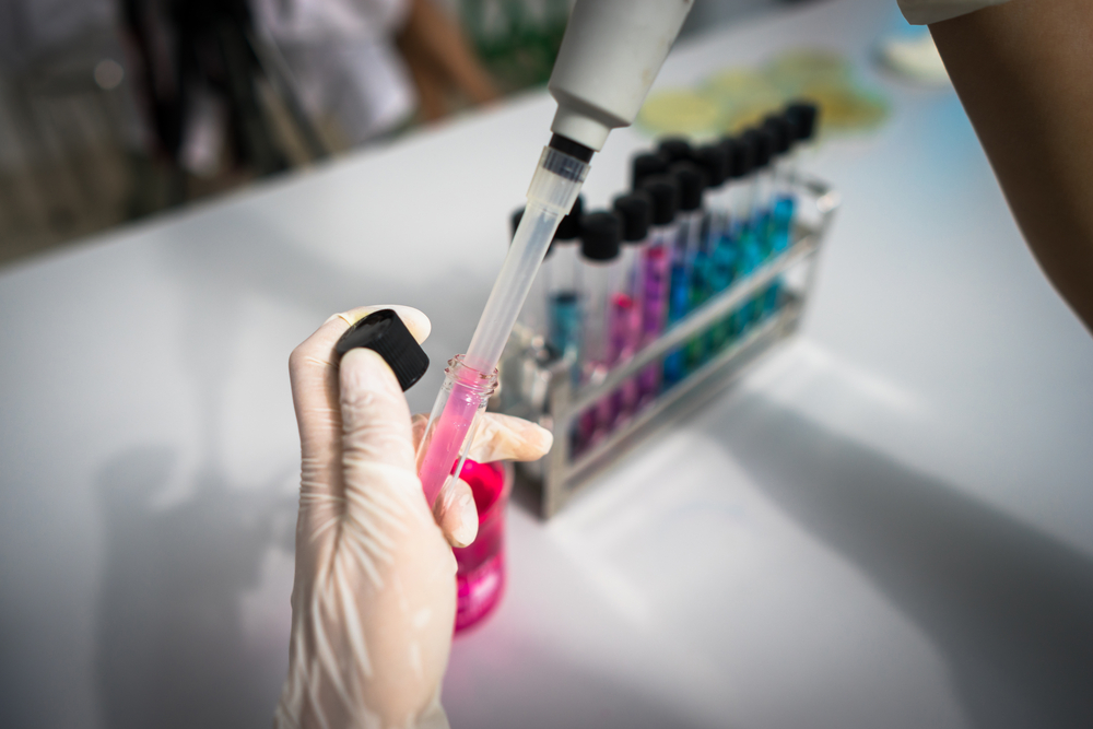
The technique centers on directly altering the biochemical signature of donor kidneys before transplantation. Blood type differences are determined by specific carbohydrate structures attached to proteins and lipids on the surface of cells. In type A kidneys, these structures include terminal sugar molecules that act as identifiers for the immune system. The research team applied a set of enzymes that selectively cleave these terminal sugars, effectively removing the defining features of the A antigen without disrupting the underlying tissue structure. This approach builds on earlier enzyme discovery work but extends it to intact human organs rather than isolated cells or laboratory samples.
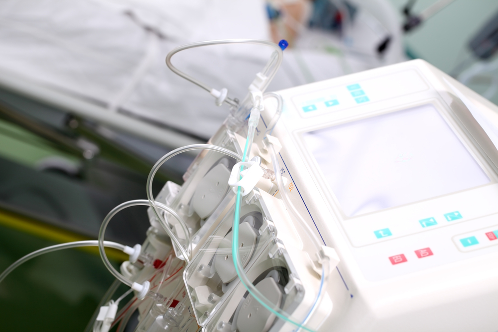
A key part of the method is how the enzymes are delivered. The kidney is connected to a perfusion system that allows fluids to circulate through its blood vessels outside the body. This setup ensures that the enzymes reach the entire organ evenly, including the small vessels where immune reactions are most likely to begin. By controlling factors such as flow rate, temperature, and exposure time, researchers can optimize how thoroughly the antigens are removed while preserving organ function. This level of control is critical because incomplete removal could still trigger an immune response after transplantation.
Biochemist Stephen Withers of the University of British Columbia explained the concept when the study was published. “It’s like removing the red paint from a car and uncovering the neutral primer,” Withers said. “Once that’s done, the immune system no longer sees the organ as foreign.” The results suggest that targeted enzymatic modification can reduce the biological signals that define blood type at the organ level, offering a practical route to expanding compatibility without changing the recipient’s immune system.
Testing the Modified Kidney
To test whether the enzyme-treated organ could function in a realistic setting, the researchers moved beyond laboratory analysis and used a brain-dead human recipient whose family had consented to the study. That model gave the team a way to study the kidney in a living human body with active circulation, organ-level physiology, and immune surveillance, while avoiding the ethical risk of exposing a living transplant patient to an experimental procedure that had not yet been proven safe. In transplant research, this kind of model can provide a more meaningful bridge between bench science and future clinical trials because it reveals how an organ performs under conditions that cannot be fully reproduced in cell studies or ex vivo testing.

The kidney survived and functioned for several days after transplantation, which allowed the researchers to monitor whether the organ could remain viable after enzymatic modification and surgical implantation. That mattered because a promising biochemical idea still has to prove that the organ can tolerate the treatment process itself, maintain blood flow, and continue performing basic kidney functions once placed in the body. According to Withers, this stage of the research provided an important milestone. “This is the first time we’ve seen this play out in a human model,” he said.
The experiment also gave the team a chance to collect information that will shape the next phase of the work, including how to refine the treatment protocol and what signals to track in later studies. Rather than answering only whether the technique can remove antigens, this stage asked a more practical question: can a modified kidney behave like a transplantable organ under human physiological conditions? “It gives us invaluable insight into how to improve long-term outcomes,” Withers added.
What Happened After the Transplant
The post-transplant period gave researchers a closer look at how stable the antigen removal process is once the kidney is exposed to a human biological environment. By the third day, the reappearance of type A markers suggested that the enzymatic modification was not fully durable under physiological conditions. This indicates that either some antigen structures were not completely removed during treatment or that underlying cellular processes were able to restore these markers over time. Understanding this reversal is important because even partial re-expression can reintroduce immune recognition.
Despite this, the immune response that followed was not as aggressive as typically seen in incompatible transplants. This suggests that reducing antigen levels, even without fully eliminating them, may be enough to blunt the intensity of early immune activation. In practical terms, this could mean that antigen reduction lowers the threshold at which the immune system reacts, which may create a window where the body is more likely to adapt to the transplanted organ rather than reject it immediately.
Researchers also observed signs consistent with early immune tolerance, where the immune system does not fully attack the transplanted tissue despite recognizing some level of difference. This is a key area of interest in transplant medicine because long-term graft survival often depends on achieving a balance between immune control and acceptance. The findings from this stage point to a shift from an all-or-nothing model of compatibility toward a gradient, where reducing incompatibility may be enough to improve outcomes, even if complete compatibility is not yet achieved.
Challenges Still Ahead
The main challenge now is translation into real-world transplant practice. An enzyme treatment that works under experimental conditions must also perform consistently across donor kidneys that vary in age, quality, ischemic time, and baseline injury. Researchers will need to show that the process can be standardized so that antigen removal is reproducible from one organ to the next without impairing the kidney’s structure, vascular integrity, or immediate post-transplant function. That is especially important in deceased donor transplantation, where timing is limited, and any added step must fit within a narrow clinical window.

There is also the question of how this method would fit into the broader transplant workflow once it reaches patient studies. Success will depend not only on whether the organ can be modified, but also on whether transplant centers can verify the extent of antigen removal before surgery, monitor for rebound expression after implantation, and integrate the method with existing preservation and immunosuppression protocols. In other words, the remaining barriers are not only biological but operational. The technique has to be reliable enough for routine use, measurable enough for clinicians to trust, and practical enough to deploy without slowing down access to transplantation.
From Lab Discovery to Real World Impact
This research reflects a long progression from basic biochemical discovery to a point where direct clinical application is within reach. Scientists first identified enzymes capable of removing blood group antigens at a molecular level, then adapted that knowledge to work on whole human organs under conditions that preserve function. What began as a question in enzyme chemistry has now advanced into a method that can be tested in human models, showing that organ-level modification is not only theoretically possible but technically achievable.
Kidney transplantation remains the most effective treatment for advanced kidney failure, yet access continues to be limited by compatibility constraints. This approach does not eliminate those barriers yet, but it introduces a practical path toward expanding who can receive a given organ. If future studies confirm that antigen removal can be made stable and reliable, the result would be a measurable increase in usable donor kidneys without requiring more donors. That shift would move transplant medicine toward a system where compatibility is engineered rather than waiting for, with direct consequences for how many patients can be treated in time.


