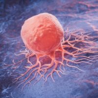
What if scientists could use a chemical produced inside a cancer cell to manufacture a compound capable of damaging it? Researchers in Japan have demonstrated an experimental technique that does precisely this in laboratory-grown cells, opening a new avenue for developing targeted cancer treatments.
The study, published in Nature Communications on September 17, 2026, uses carbon monoxide as a chemical building block to produce potentially cancer-killing compounds inside cells. The results are intriguing, but the researchers observed only modest anticancer effects, and the technique has not been established as a treatment for patients.
How Scientists Turned Cancer Cells Into Chemical Factories
The research team, which included scientists from RIKEN and the Institute of Science Tokyo, developed a system that takes advantage of carbon monoxide produced naturally inside certain cancer cells. Rather than simply delivering a finished drug, the researchers designed a way to manufacture an active compound within the cell using its existing chemical environment.
Carbon monoxide is commonly associated with poisoning because it interferes with the body’s ability to transport oxygen. However, the body also produces small amounts of this gas naturally, where it serves biological signalling functions. Studies have found elevated carbon monoxide production in certain cancer cells, offering researchers a possible chemical feature to exploit.
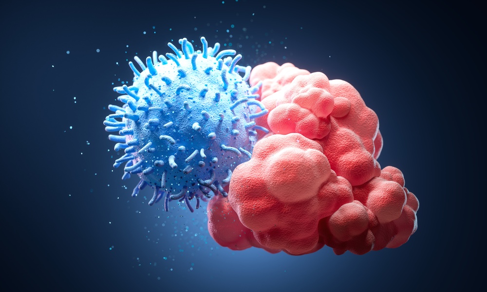
The team, led by Katsunori Tanaka, developed an albumin-palladium complex to make use of this difference. Their system combines a protein that can carry molecules with a metal-based catalyst that helps drive the chemical reaction. When the components enter a suitable cellular environment, the catalyst can use available carbon monoxide to help produce phenanthridinone derivatives.
Some members of this compound family have biological activities relevant to cancer research. The researchers therefore investigated whether their delivery system could generate an active compound inside cancer cells and suppress their growth. Their findings support the feasibility of this approach under laboratory conditions, although much more work is needed before its potential medical value can be established.
What Makes the Experimental Technique Different?

Many cancer treatments involve administering a drug that travels through the body before reaching its target. Although targeted therapies can improve treatment precision, delivering enough of a drug to a tumour while limiting its effects on healthy tissues remains a major challenge.
The Japanese researchers explored another strategy by combining drug synthesis with delivery. Their system attempts to produce an active compound after entering a cell, using carbon monoxide already present in that environment as a building block. This approach could eventually offer an additional way to control where certain therapeutic compounds are generated.
The Role of Albumin
Albumin is a protein naturally found in blood and commonly studied for its ability to carry different substances. In this experiment, the researchers used albumin as a protective structure for their palladium-based catalyst, placing the metal complex within a hydrophobic pocket of the protein.
This protection addresses a significant chemical obstacle. Inside cells, a molecule called glutathione can deactivate certain metal catalysts, preventing them from completing the reactions needed to produce a drug. By shielding the palladium complex within albumin, the researchers helped preserve its ability to react under conditions that would otherwise interfere with its activity.
The albumin structure also helped the system enter cells through a process known as endocytosis, in which cells take up material from their surroundings. Once inside, the complex could participate in the chemical reactions required for producing the desired compounds.
How Carbon Monoxide Helps Produce the Compound
Carbon monoxide acts as a raw material in the reaction rather than simply serving as a signal that identifies a cell. The palladium catalyst helps incorporate it into the chemical structure of phenanthridinone derivatives, some of which have properties that researchers are investigating for cancer treatment.
The team also developed a release-type system designed to allow the newly produced active compound to separate from the delivery structure. This is important because the resulting molecule needs to be available to act inside the cell rather than remaining attached to the protein-bound components.
The process represents a specialised form of chemical synthesis inside a biological system. It demonstrates that certain therapeutic molecules can be assembled within laboratory-grown cells, although the same process must still be evaluated in the more complex conditions of a living organism.
What the Laboratory Experiments Revealed

The researchers tested their approach across several stages, first examining whether the protected catalyst could produce the intended reaction products. They then investigated whether the system could generate an active compound inside cancer cells and affect their ability to grow.
Their experiments included human cancer cell lines derived from liver, breast, lung and cervical cancers. The team detected the desired chemical products in the tested cancer cells, providing evidence that the reaction could occur in more than one laboratory model.
The researchers subsequently developed a system designed to produce an oxynitidine-derived phenanthridinone compound with cytotoxic activity. Cytotoxicity refers to the ability of a substance to damage or kill cells, which can be useful in cancer treatment but can also create risks if healthy cells are affected.
In liver cancer cells, the team detected the production of the active compound and observed effects on cell growth. Additional experiments involving breast cancer cells found that the treatment system could suppress colony formation, a measure used to assess whether cells retain the ability to grow and reproduce over time.
These results offer evidence that the technique can do more than detect carbon monoxide. Under the conditions tested, it could also use the molecule to help produce an active compound with an effect on cancer cell growth. However, the experiments do not establish that the technique can eliminate tumours or improve outcomes for people with cancer.
Did the Technique Spare Healthy Cells?
One of the most important questions in cancer treatment is whether a therapy can damage cancer cells while limiting harm to healthy tissue. The researchers investigated this issue by comparing their experimental system’s effects on cancer cells with its effects on non-cancerous fibroblasts.
Fibroblasts are connective-tissue cells involved in maintaining and repairing tissues throughout the body. They provide a useful comparison in laboratory experiments, but they cannot represent every type of healthy cell that a treatment might encounter in a person.
The study found evidence of reduced toxicity in normal fibroblasts compared with the liver cancer cells tested under certain conditions. The researchers also reported that the observed anticancer effect was modest, meaning the results did not demonstrate a powerful or complete elimination of cancer cells.
These distinctions are important because the presence of a potential difference between cancerous and non-cancerous cells does not automatically establish a safe treatment window. A future therapy would need to show that it can produce a sufficiently strong effect against cancer while keeping damage to healthy tissues within acceptable limits.
Why Carbon Monoxide Levels Matter
The proposed strategy depends on the availability of carbon monoxide inside the target cell. If the relevant chemical conditions differ substantially between cancer cells and healthy cells, the system might produce different amounts of the active compound. However, cancer is biologically diverse, and researchers cannot assume that every tumour will have the same chemical characteristics.
The researchers also considered the possibility of premature drug production in the bloodstream. Most carbon monoxide circulating in blood is bound to haemoglobin, the oxygen-carrying protein in red blood cells, which makes it less available for the reaction. Their findings support the idea that the system may respond preferentially to available carbon monoxide inside certain cancer cells, but this does not prove that premature activation or other safety problems would never occur in a living body.
The selectivity of the technique must therefore be investigated further. Researchers need to establish how consistently it distinguishes cancer cells from healthy tissue and whether its activity changes across different tumour types.
Why This Research Could Matter for Future Treatments
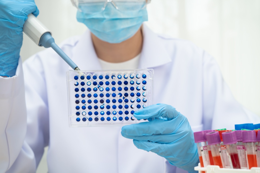
The study explores an approach that could eventually complement existing strategies for developing cancer medicines. By using a molecule associated with certain cancer cells as a chemical building block, researchers are investigating whether the local environment of a tumour can help determine where an active compound is produced.
If this principle can be developed successfully, it could provide another way to control drug production within diseased tissue. Generating an active compound near its intended target might help reduce exposure elsewhere in the body, although that benefit has not yet been demonstrated in patients using this particular system.
The research also has implications for the broader field of therapeutic in vivo synthetic chemistry. This area investigates whether chemical reactions can be performed within living biological systems to create useful molecules directly where they are needed. The Japanese team’s work advances that concept by demonstrating the use of an internally produced molecule in the synthesis of potentially therapeutic compounds.
Several challenges must still be addressed before this strategy can become a practical treatment. Researchers need to establish whether the system can reach tumours effectively, remain active under real biological conditions and generate enough of the intended compound to produce a meaningful therapeutic effect.
They must also investigate how the body handles the albumin-palladium complex, whether the components accumulate in particular organs and what side effects might occur. These questions cannot be answered by examining cultured cells alone, because drug distribution, metabolism and immune responses can all influence how a treatment behaves in a living organism.
What Scientists Still Need to Establish
The next stage of research must determine whether the laboratory findings can be reproduced and extended beyond the cellular models used in the study. Evidence from additional experiments will be needed to establish the technique’s safety, effectiveness and suitability for further development.
Several questions remain central to that process:
- Effectiveness in living organisms: Researchers need to determine whether the system can reach tumours and suppress their growth in animal models before its potential for clinical testing can be assessed.
- Safety for healthy tissues: Further testing must establish whether the catalyst, delivery structure and active compound cause harmful effects in organs or other non-cancerous cells.
- Differences between tumours: The technique depends on cellular chemistry, so scientists must investigate whether its performance varies between cancer types and individual tumours.
- The strength of the treatment effect: The observed anticancer activity was modest, making it important to determine whether the system can be improved enough to produce a meaningful therapeutic benefit.
- Reliable drug production: Researchers must establish whether the chemical reaction can occur consistently under biological conditions and whether the amount of compound produced is sufficient to affect a tumour.
These are substantial research challenges, and success in one area would not automatically resolve the others. Even if future studies demonstrate encouraging results in animals, further testing would still be necessary before researchers could determine whether the approach is safe and effective for people.
What This Means for People Living With Cancer
The findings provide a useful example of how researchers are exploring new ways to address the difficulties of cancer treatment. Instead of relying only on drugs manufactured in advance, the team demonstrated a method for producing an active compound inside laboratory-grown cancer cells by using a molecule already present in those cells.
However, the results should not be interpreted as evidence that cancer can now be made to destroy itself in patients. The experiments established a chemical proof of concept, and the observed effects were modest. The study does not demonstrate a cure, establish improved survival or show that the technique is ready for medical use.
For anyone affected by cancer, it is important to distinguish experimental research from treatments supported by clinical evidence. Decisions about treatment should continue to be based on established medical guidance and individual clinical circumstances, rather than expectations created by early laboratory findings.
The most meaningful next development would be evidence that the technique can safely reach tumours in living organisms, produce a strong enough anticancer effect and limit harm to healthy tissue. Until those results are available, the Japanese researchers’ work remains an interesting scientific advance with potential that still needs to be tested.