
Imagine doctors being able to deliver specialized materials directly to damaged tissue deep inside the body, without needing to reach the area through traditional surgical procedures. Scientists have taken a significant step toward making this possible after developing a technique that uses focused sound waves to trigger 3D printing inside living animals. The technology could eventually help deliver medication, support tissue repair, and monitor internal organs with greater precision.
The research, led by scientists at the California Institute of Technology (Caltech), has already produced some interesting results in mice. Using a technique called deep tissue in vivo sound printing (DISP), researchers successfully created drug-loaded polymer structures near bladder tumors and observed substantially more tumor cell death than in animals receiving the drug through direct injection. The technology is still experimental, but its potential applications could extend far beyond cancer treatment.
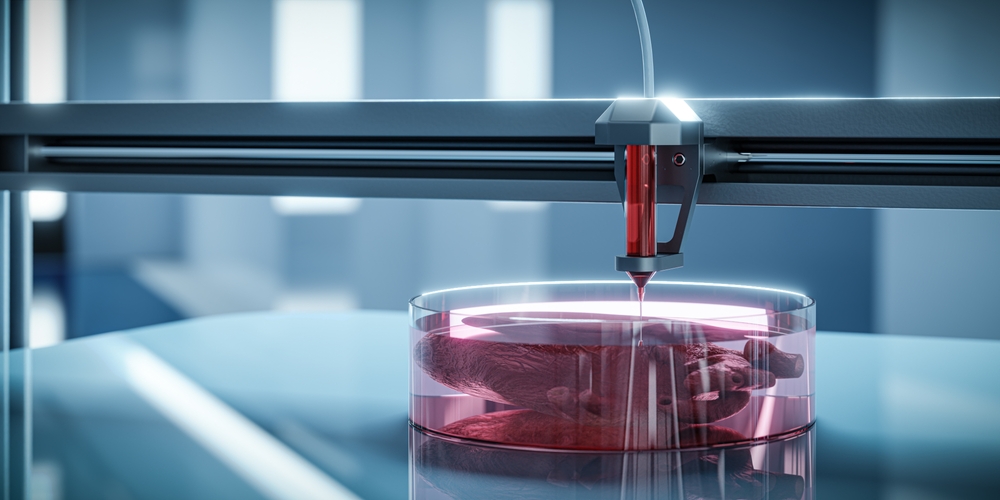
Scientists Find A Way To Print Deep Inside Living Tissue
The research team developed a method that allows polymers to form at specific locations inside living animals using ultrasound. Polymers are materials made from repeating molecular building blocks, and they can be designed to carry drugs, support tissue repair, or perform other medical functions. The challenge has always been controlling exactly where these materials form when they are introduced into the body.
Previous approaches used infrared light to trigger polymerization, the process through which individual building blocks connect to form a larger material. However, infrared light has limited penetration through biological tissue, making it difficult to reach deeper areas of the body. Wei Gao, professor of medical engineering at Caltech and a Heritage Medical Research Institute Investigator, explained that limitation: “But infrared penetration is very limited. It only reaches right below the skin.”
The new approach uses ultrasound because sound waves can penetrate much deeper into biological tissue. Gao explained that the technique can reach deep tissue while allowing researchers to print different materials with good biocompatibility. The study was published in the journal Science, with Elham Davoodi, an assistant professor of mechanical engineering at the University of Utah, serving as its lead author. Davoodi completed the research while working as a postdoctoral scholar at Caltech.
How Sound Waves Trigger The 3D Printing Process
The technology combines focused ultrasound with microscopic structures called liposomes. These spherical particles have protective fatty layers and are already widely used in drug delivery research because they can carry different substances. For this experiment, scientists loaded the liposomes with a chemical agent that triggers polymerization, then incorporated them into a specially prepared liquid mixture containing the materials needed for printing.
This mixture is known as bioink. It contains the basic building blocks of the desired polymer, the specially prepared liposomes, an imaging contrast agent, and whatever therapeutic material researchers want to deliver. Depending on the intended application, the mixture can also contain living cells or conductive substances such as carbon nanotubes and silver. Once prepared, the bioink is injected directly into the body, where researchers can use focused ultrasound to activate it at a selected location.
The key is that the liposomes respond to small temperature changes. By directing ultrasound toward a particular area, researchers can increase its temperature by approximately 5 degrees Celsius. This causes the liposomes to release their chemical contents, triggering the polymer building blocks to connect and form a solid material. Gao explained: “Where the agents are released, that’s where localized polymerization or printing will happen.” This allows the researchers to control where the printing takes place instead of having the entire injected mixture solidify.
Researchers Can Monitor Where The Material Forms
Controlling the printing location is only one part of the process. Scientists also need to know whether the material has formed correctly and whether it has appeared in the intended area. To achieve this, the research team incorporated gas vesicles derived from bacteria into the bioink. These tiny protein structures contain air and produce a strong signal during ultrasound imaging, making them useful for observing changes inside tissue.
The gas vesicles respond to chemical changes that occur when the liquid monomer solution transforms into a gel network. Their ultrasound imaging contrast changes during this transformation, allowing researchers to identify where polymerization has taken place. This gives the team a way to monitor the printing process and determine the location of the newly formed material while working inside living animals.
The imaging component could become particularly useful for applications involving organs that are difficult to access. If researchers can identify precisely where a polymer forms, they have greater control over how therapeutic materials are delivered. This combination of ultrasound activation and imaging also gives the technology an advantage over approaches that depend on light, which cannot easily reach deep internal structures.
The First Tests Showed Promising Results Against Tumors
One of the most significant demonstrations involved mice with bladder tumors. Researchers used the DISP platform to print polymer structures loaded with doxorubicin, a chemotherapy drug, near the tumors. They then compared the results with animals that received the medication through direct injection of drug solutions.
The mice treated using the drug-loaded printed hydrogels experienced substantially more tumor cell death over several days. This suggests that creating a drug-containing material near a tumor could influence how effectively medication reaches the intended area. The findings provide an early demonstration of how the platform might eventually support more targeted drug delivery.
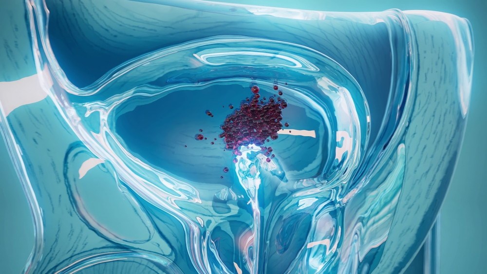
Doxorubicin is a chemotherapy medication used in cancer treatment, but chemotherapy can also affect healthy cells. Researchers are therefore interested in approaches that could concentrate treatment closer to diseased tissue. Gao explained that the team had already demonstrated drug-loaded hydrogel printing for tumor treatment in a small animal model. However, the results do not establish that the technology is ready for human cancer treatment, and further research will be needed to assess its safety and effectiveness.
The Technology Could Eventually Support Tissue Repair
Cancer treatment is only one possible application of the new technique. Researchers have also demonstrated the ability to print bioadhesive gels and polymers that could help seal internal wounds. These materials have glue-like properties and could eventually provide another way to address injuries inside the body.
Another potential application involves delivering cells to damaged tissue. Imagine a future treatment designed to place specialized cells exactly where they are needed to support tissue repair. The researchers believe the platform could eventually help make this possible by allowing cells to be incorporated into the bioink before it is injected. One example explored in the research’s broader vision involves delivering materials that could support tissue repair inside a damaged heart.
The team has also demonstrated the printing of bioelectric hydrogels. These are polymer materials containing conductive components that could be used for monitoring physiological signals inside the body. The researchers describe potential applications involving internal monitoring of vital signs, including signals associated with electrocardiograms (ECGs). These developments could eventually contribute to new approaches for monitoring organ activity, although further testing will be needed before such applications become practical medical treatments.
Artificial Intelligence Could Help Print Inside A Beating Heart
One of the biggest challenges facing the technology is movement inside the body. Some organs constantly change position and shape as they perform their normal functions. The heart is a particularly difficult example because it contracts and relaxes continuously, making it challenging to direct a printing process toward one precise location.
The researchers believe machine learning could help improve the accuracy of the system. Artificial intelligence could potentially assist with identifying target locations and adjusting focused ultrasound as organs move. This would be especially useful if scientists eventually want to create polymer structures inside a beating heart without losing precision during the printing process.
Gao described the team’s ambition: “In the future, with the help of AI, we would like to be able to autonomously trigger high-precision printing within a moving organ such as a beating heart.” The researchers are now interested in testing the technology in larger animal models. Gao also expressed hope that human evaluation could become possible in the future, although no human application has yet been established.
What This Could Mean For Future Medical Treatments
The DISP platform brings together ultrasound, temperature-sensitive liposomes, imaging technology, and specially designed polymers to create materials inside living animals. Each component performs a different role, from activating the printing process to showing researchers where the material has formed. Combining these functions gives scientists a new way to investigate targeted medical treatments.
Potential applications being explored include:
- Targeted drug delivery: Printing medication-loaded hydrogels near tumors or other affected tissue.
- Tissue repair: Delivering specialized materials and potentially therapeutic cells to damaged areas.
- Internal wound sealing: Creating bioadhesive polymers that could help close internal injuries.
- Heart monitoring: Printing conductive hydrogels that could support the monitoring of physiological signals.
- Precision treatment: Using imaging and focused ultrasound to control where materials form inside the body.
There are still important questions about how safely these materials can be used over longer periods, how the technology will perform in larger organisms, and whether the same precision can be achieved in humans. The current findings come from animal research, so they should not be interpreted as evidence that doctors can already perform internal 3D printing as a routine medical procedure.
A New Direction For Medicine Is Taking Shape
The ability to create specialized materials deep inside living tissue could eventually change how researchers approach certain medical problems. Instead of relying entirely on conventional methods to reach difficult areas, doctors may one day have access to technologies that activate therapeutic materials from outside the body. The Caltech team’s findings offer an early demonstration of that possibility, particularly through its experiments involving drug delivery near bladder tumors in mice.
For now, the next major milestone is testing the technology in larger animals and determining whether it can meet the safety and precision requirements of human medicine. The prospect of printing therapeutic materials inside a moving organ remains a future research goal, but scientists have already demonstrated that sound waves can do something once considered extremely difficult: trigger controlled material formation deep inside a living body.
Sources:
Davoodi, E., Li, J., Ma, X., Najafabadi, A. H., Yoo, J., Lu, G., Sani, E. S., Lee, S., Montazerian, H., Kim, G., Williams, J., Yang, J. W., Zeng, Y., Li, L. S., Jin, Z., Sadri, B., Nia, S. S., Wang, L. V., Hsiai, T. K., . . . Gao, W. (2025b). Imaging-guided deep tissue in vivo sound printing. Science, 388(6747), 616–623. https://doi.org/10.1126/science.adt0293
Davoodi, E. (2026, May 23). Imaging-guided deep tissue in vivo sound printing. Davoodi Lab. https://davoodilab.mech.utah.edu/2025/05/10/imaging-guided-deep-tissue-in-vivo-sound-printing/