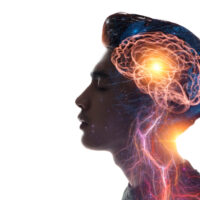
A discovery that began with a single-celled alga has now earned the 2026 Nobel Prize in Physiology or Medicine. The breakthrough gives researchers an extraordinary way to switch specific nerve cells on and off using light, allowing them to study the brain with a level of precision that was previously out of reach.
The technique, known as optogenetics, is already helping scientists investigate memory, behavior, pain and neurological disorders. Researchers are also testing whether it could restore limited vision in people with certain forms of blindness, offering a glimpse at how a basic biological discovery might eventually influence treatments for serious diseases.
Three Scientists Won The 2026 Medicine Nobel
Karl Deisseroth of Stanford University, Peter Hegemann of Humboldt University of Berlin and Georg Nagel of the University of Würzburg received the 2026 Nobel Prize in Physiology or Medicine for discoveries that established the foundations of optogenetics. The Nobel Assembly recognized their work on light-gated ion channels and the development of methods that allow researchers to control selected nerve cells with light.
Neuroscientist Michael Häusser of University College London called the award “a fantastic recognition of a transformative discovery that has changed the way we do neuroscience and also promises to change the way we treat diseases in the brain.” The recognition reflects how dramatically optogenetics has changed the study of the nervous system since the technique emerged in the 2000s.
For decades, neuroscientists could observe which areas of the brain became active during particular behaviors. They could also damage or stimulate regions and examine what happened afterward, but those approaches often made it difficult to establish precise cause and effect because many surrounding cells could be affected at the same time.
The goal was to develop something closer to a biological switch that could control particular neurons without disturbing nearby cells. Abdel El Manira, a neuroscientist and member of the Nobel Committee, described the need for such a tool during the award announcement, explaining that neuroscientists had dreamed of a neuronal switch that could turn specific neurons on or off with great precision.
The Story Began With A Single-Celled Alga
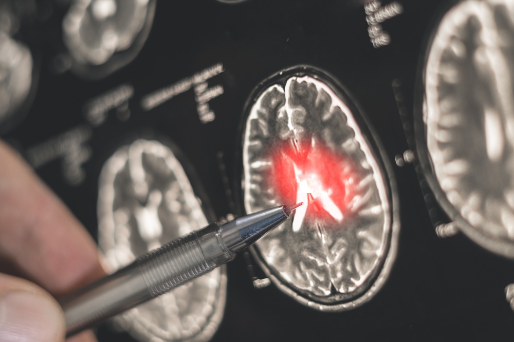
The origin of this Nobel-winning technology is surprisingly distant from the human brain. Peter Hegemann and Georg Nagel were studying how a tiny green alga called Chlamydomonas senses light and moves toward a light source, a basic biological behavior that eventually provided scientists with a way to manipulate nerve cells.
In the early 2000s, Hegemann and Nagel discovered a light-sensitive protein called channelrhodopsin on the surface of the alga’s cells. When blue light reaches the protein, a channel opens and allows positively charged ions to flow into the cell, creating an electrical impulse that helps the organism respond to light.
The researchers discovered that channelrhodopsin had a property that made it particularly valuable for neuroscience. When the protein was introduced into other types of cells, those cells could also become responsive to light, suggesting that the protein could potentially be used as a biological tool rather than simply studied as part of algae.
That finding caught Karl Deisseroth’s attention as he was developing his own research program at Stanford. He wanted a way to trigger electrical signals in individual groups of neurons, and channelrhodopsin offered a possible route to doing that with much greater precision than existing techniques.
How Light Can Control A Nerve Cell
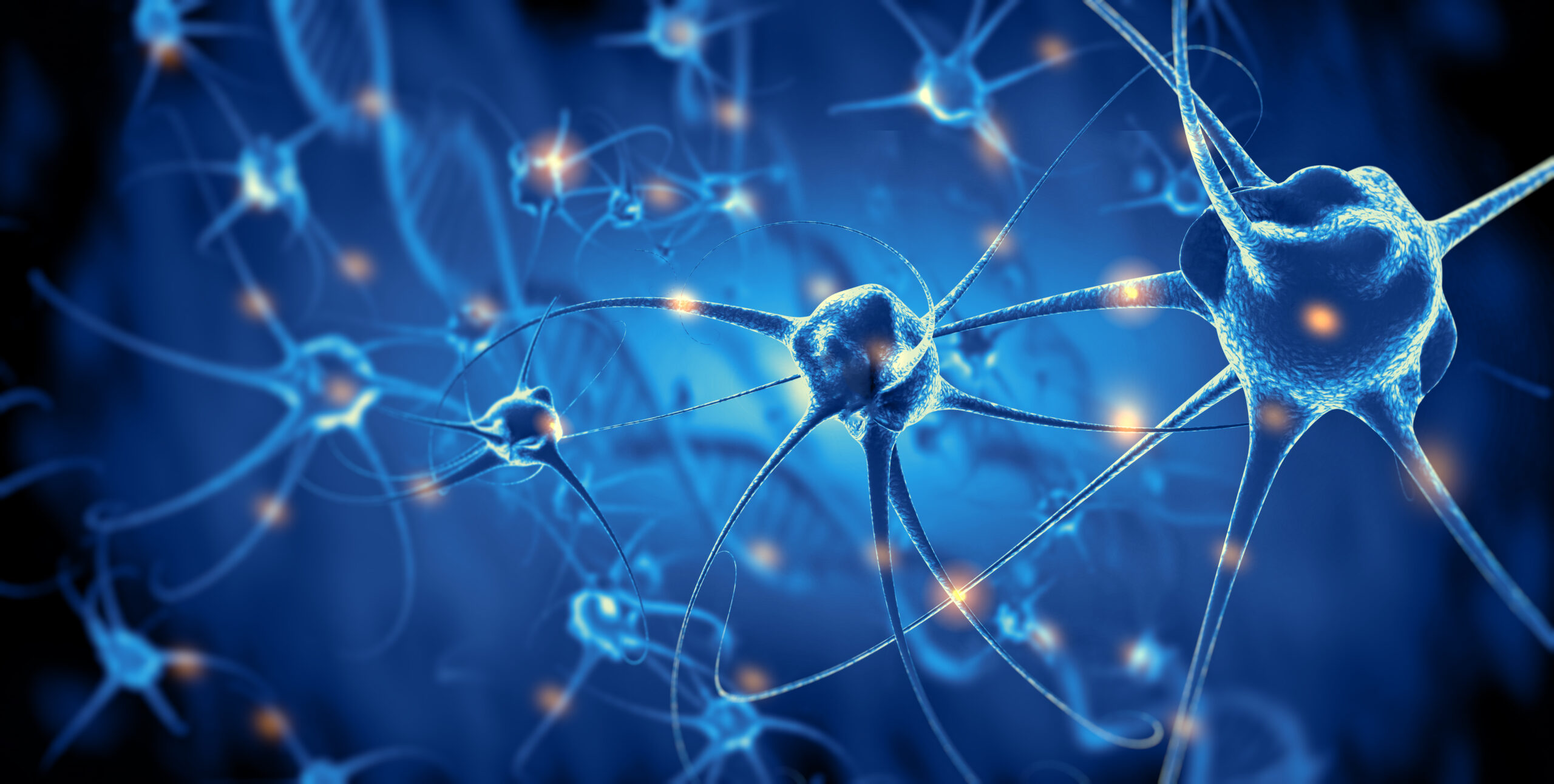
Deisseroth obtained the DNA encoding channelrhodopsin and introduced its gene into rat neurons grown in a laboratory dish. When blue light was directed at those modified neurons, the cells responded by producing a nerve signal, showing that the light-sensitive protein could function inside neurons as well as in algae.
The breakthrough became even more significant when Deisseroth and his colleagues adapted the approach for living animals. They used a thin optical fiber to deliver light to targeted neurons inside the brains of mice, allowing researchers to activate specific cells while observing what happened to the animal.
Deisseroth later described the approach as a risky choice among the methods he was considering. “It was the approach with the highest risk, but it turned out to be the one that worked best, and that was a valuable lesson,” he said in a statement.
The basic process can now be summarized in several stages:
- Targeted neurons: Researchers identify the particular group of nerve cells they want to study and introduce a gene that causes those cells to produce a light-sensitive protein.
- Light delivery: A very thin optical fiber can deliver light to the selected area of the brain in laboratory animals, allowing researchers to control when the modified cells are activated.
- Neural response: When the appropriate wavelength of light reaches the protein, the protein changes the electrical activity of the neuron.
- Behavioral observation: Scientists can then observe what happens when those cells are activated or silenced, helping them connect specific neural activity with behavior or other brain functions.
This precision is what makes optogenetics so different from older electrical stimulation methods. Andrew Jackson of Newcastle University compared earlier stimulation techniques to “playing a piano with a mallet,” because electrical stimulation could affect nearby neurons as well as the intended cells.
Scientists Can Now Test Brain Connections Directly
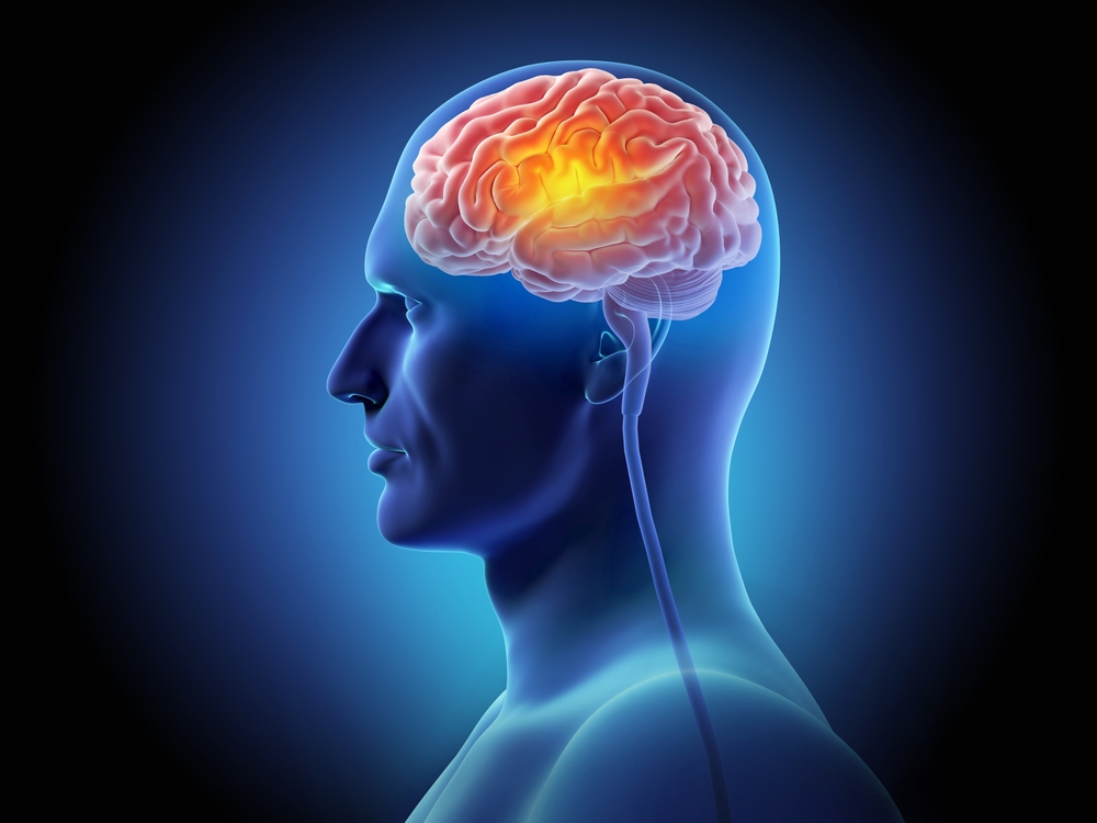
The brain contains vast networks of interconnected neurons, and many behaviors depend on several circuits working together. Researchers have long been able to observe patterns of activity, but observing a pattern does not necessarily prove that the activity caused the behavior.
Optogenetics provides a way to manipulate selected cells and then measure the consequences. That makes it possible to investigate whether a particular population of neurons is actually responsible for a memory, movement, sensation or behavioral response.
Researchers have used the method to study circuits associated with memory, pain, reward, movement and other aspects of cognition and behavior. The technology can also be adapted to target particular cell types, giving scientists a more detailed picture of how different populations contribute to the brain’s functions.
That distinction is important when studying neurological and psychiatric conditions. A disorder may involve changes across complicated neural networks, and knowing which cells contribute to a symptom could eventually help researchers identify more precise treatment targets.
Michael Häusser described optogenetics as having “completely revolutionized how we probe neural circuits in neuroscience.” The technology has also helped inspire related approaches that seek to control specific neural pathways without relying on conventional electrical stimulation.
One Experiment Showed How Memory Could Be Manipulated
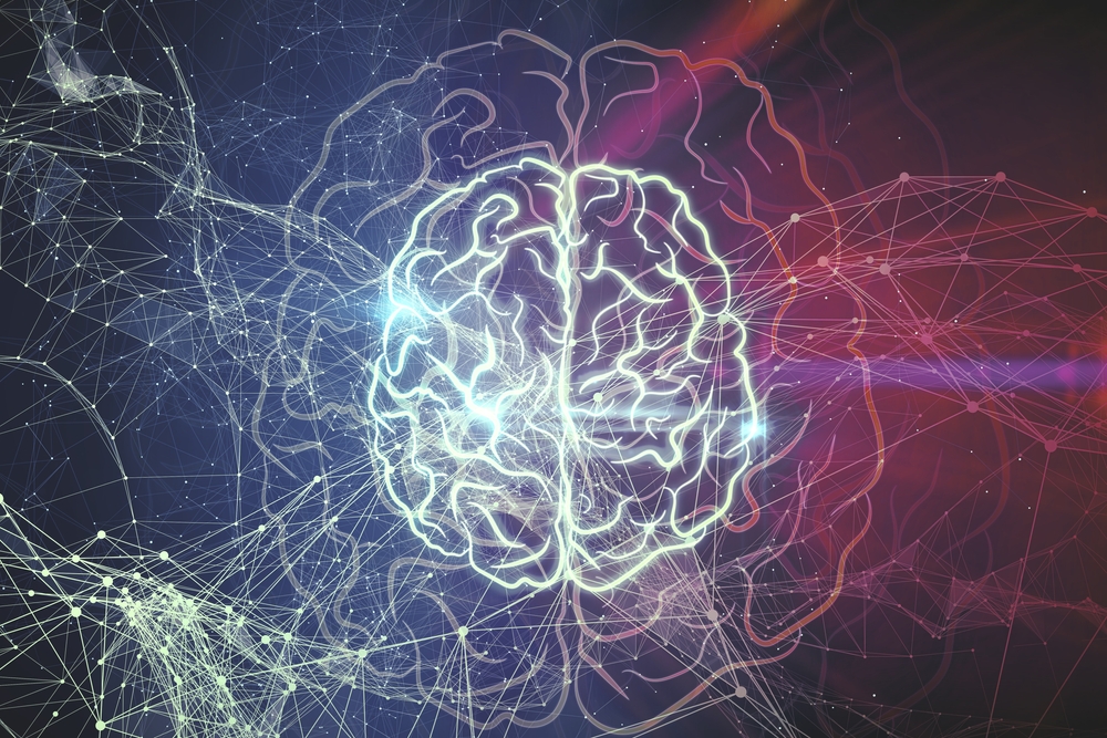
One of the most striking demonstrations of optogenetics involved memory and fear in mice. In a 2012 experiment, researchers exposed mice to a mild electric shock in a particular cage and used optogenetics to identify neurons in the brain’s memory center that were active during the experience.
Several days later, the mice were placed in a different cage that did not carry the same context. Researchers then used light to activate the neurons that had been associated with the earlier experience, and the mice responded by freezing, a behavior associated with fear.
The experiment gave researchers a powerful way to investigate the connection between defined neurons and a particular memory. Instead of simply watching which cells became active during an experience, scientists could manipulate the cells and observe whether activating them produced a related response.
That approach has helped move neuroscience toward more precise questions about how memories are stored and recalled. It also illustrates why the Nobel-winning technique has attracted so much attention from researchers studying conditions in which memory, emotion and behavior become disrupted.
The work does not mean scientists can simply switch human memories on and off. Most of these experiments have been conducted in animals, and translating precise neural manipulation into safe human treatments presents major scientific and technical challenges.
Optogenetics Is Already Being Tested For Vision Loss

One of the most significant steps toward human applications has involved retinitis pigmentosa, a genetic condition in which light-sensitive cells in the retina gradually deteriorate. As those photoreceptor cells are lost, the eye becomes less capable of converting light into the electrical signals normally sent to the brain.
Researchers are investigating whether optogenetics can bypass some of that damage by making surviving retinal nerve cells sensitive to light. The approach involves introducing a gene for a light-sensitive protein into cells that remain in the retina after the normal photoreceptors have been lost.
The modified cells can then respond to light, creating a potential route for sending visual information toward the brain. The method does not recreate a healthy retina, but it could provide some visual function when the original light-detecting cells are no longer available.
In 2021, researchers reported using an optogenetic approach in a blind man with retinitis pigmentosa. With the help of light-amplifying goggles, he was able to make out objects, demonstrating that the basic concept could produce a measurable visual response in a person.
The result was limited, and it should not be confused with a complete restoration of normal eyesight. Still, the experiment showed that researchers could use genetic modification and light-sensitive proteins to create a new route for visual information in an eye affected by retinal degeneration.
Other Brain Diseases Are Being Investigated
Vision is only one area where researchers see potential applications. Optogenetic studies in laboratory animals have helped scientists examine the neural circuits involved in conditions including Parkinson’s disease, epilepsy, addiction and obsessive-compulsive disorder.
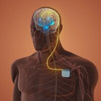
The technique has also contributed to research surrounding deep-brain stimulation, or DBS, which is already used clinically for certain neurological conditions. By studying which neural circuits are affected by stimulation, scientists hope to understand how DBS works and whether future approaches could target the relevant pathways with greater precision.
The same principle could prove useful for investigating psychiatric conditions. Disorders such as depression and addiction involve complicated interactions between different brain circuits, and identifying the specific pathways involved could help researchers develop more targeted treatments.
There is still a substantial gap between laboratory research and routine medical care. Optogenetics requires researchers to introduce light-sensitive proteins into cells, and delivering light precisely to deep regions of the human brain is technically difficult.
For that reason, some scientists are developing related technologies that could achieve similar precision without requiring a light source to reach every targeted neuron. These approaches remain areas of active research rather than established treatments.
New Technologies Could Build On The Same Idea
The influence of optogenetics can already be seen in other areas of neuroscience. Researchers have developed approaches such as chemogenetics, in which neurons are modified to carry receptors that respond to specially designed drugs.
Another developing area is sonogenetics, which explores whether certain ion channels could be activated using ultrasound. Both approaches reflect the same broader goal: finding ways to control defined groups of neurons while avoiding widespread disruption of surrounding brain tissue.
These technologies could eventually prove more practical for certain human applications. Light works extremely well in laboratory experiments because researchers can precisely control its timing and location, but delivering light deep into the human brain is considerably more complicated.
The Nobel-winning work therefore represents more than one medical technique. It helped establish a way of thinking about the brain in which individual populations of neurons can be identified, manipulated and studied as components of larger circuits.
That shift could influence future research even when optogenetics itself is not the treatment ultimately used in patients.
What The Nobel Means For Future Brain Health
The 2026 Nobel Prize does not mean that optogenetics is ready to treat most neurological diseases today. Its biggest impact remains in neuroscience research, where it has given scientists a powerful method for investigating how individual neural circuits contribute to behavior and brain function.
The medical possibilities are nevertheless becoming easier to see. Early work involving vision shows that researchers can move the technology from animal experiments toward human testing, while studies involving Parkinson’s disease, epilepsy, pain and other conditions are helping identify potential targets for future therapies.
Karl Deisseroth has said that one of the most exciting aspects of the technology is its ability to break complicated brain states into their underlying cellular components. Understanding which cells and connections produce particular symptoms could eventually help researchers design treatments around those specific pathways.
That could be especially valuable in disorders where broad treatments affect many parts of the nervous system. More precise therapies could potentially reduce unwanted effects by focusing on the circuits that actually contribute to a particular condition.
The technology also illustrates why basic science can have consequences far beyond its original purpose. Hegemann and Nagel were investigating how a microscopic alga responds to light, while Deisseroth was searching for a precise way to control neurons. Their paths eventually converged into a technology that has reshaped modern neuroscience.
For people living with neurological disease or vision loss, the most important part of this Nobel recognition may be what researchers can learn next. The treatments are still developing, but scientists now have a much sharper tool for asking which cells drive disease and how those circuits might one day be changed.
A microscopic organism helped provide the switch, and researchers are now learning how to use it to understand one of the most complicated systems in the human body. That progression from basic biology to potential medicine is exactly why this discovery could remain important long after the Nobel ceremony is over.