For people living with severe spinal cord injuries, regaining the ability to walk can seem out of reach. However, a new scientific discovery is offering a closer look at how electrical stimulation may help restore movement, with researchers identifying specific nerve cells that play a critical role in the recovery process. The findings follow a clinical trial involving nine people whose walking ability improved when spinal cord stimulation was switched on, raising new questions about how the nervous system responds to treatment.
Published in Nature, the study examined the biological mechanisms behind a technique known as epidural electrical stimulation. Researchers discovered that particular neurons in the lower spinal cord were essential for restoring walking in injured mice receiving the treatment. The findings could help scientists develop more targeted approaches to rehabilitation, although further research is needed to determine how these cellular mechanisms can be used to improve recovery in people living with paralysis.

Nine Patients Experienced Improvements In Walking Ability
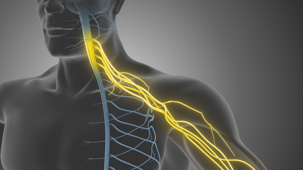
Spinal cord injuries can interrupt the communication pathways between the brain and the nerve circuits that control movement. Depending on the severity of the damage, people may experience partial or complete paralysis, making everyday activities difficult and limiting their independence. Over recent years, scientists have developed electrical stimulation systems designed to activate neural circuits below the site of an injury, helping some patients recover movement that was previously difficult or impossible.
Researchers at the Swiss Federal Institute of Technology Lausanne (EPFL), working with neurosurgeon Jocelyne Bloch at Lausanne University Hospital, investigated a technique called epidural electrical stimulation. The approach uses implanted electrodes to deliver electrical pulses to the spinal cord, helping activate neural networks involved in movement. When combined with motor rehabilitation, the technique has shown potential for improving mobility in people with severe spinal cord injuries.
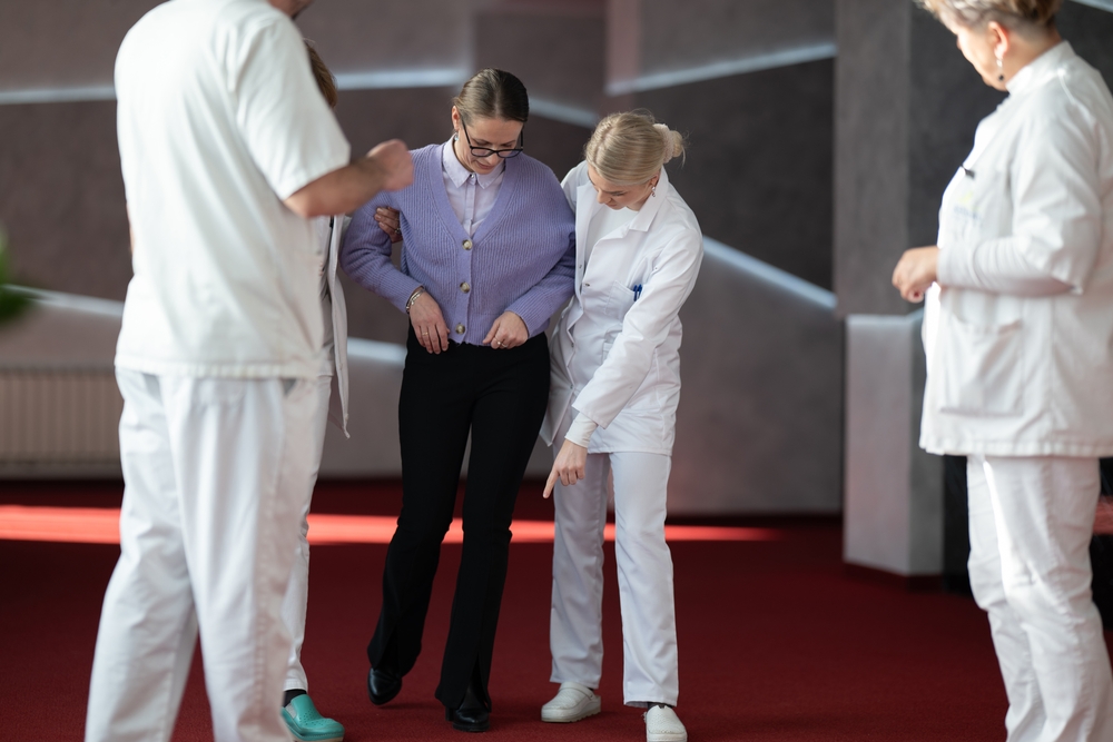
In the clinical trial described in the research, nine patients experienced immediate improvements in their walking ability when the stimulation was activated. Some participants also retained improved motor function after the stimulation was switched off. This observation caught the researchers’ attention because it suggested that the effects might extend beyond the temporary activation of neural circuits during treatment.
The possibility of lasting changes led the team to investigate what was happening inside the spinal cord. If stimulation could influence the way certain neurons functioned after an injury, understanding those cells might help scientists refine treatments and identify more precise ways to support recovery.
How Electrical Stimulation Helps The Spinal Cord
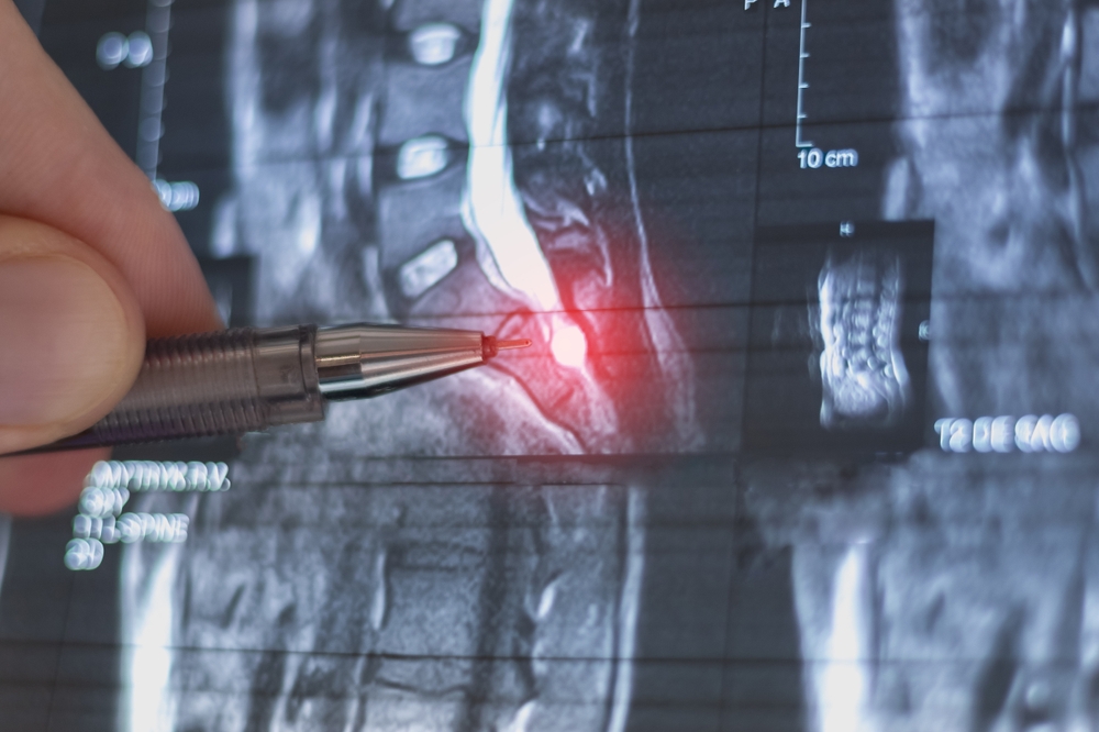
A spinal cord injury can damage the pathways that carry signals from the brain to the muscles. Even when muscles remain physically capable of movement, disrupted communication can prevent the nervous system from coordinating the movements required for walking. The spinal cord also contains neural circuits that contribute to the repeated patterns of muscle activity involved in standing and moving the legs.
Epidural electrical stimulation attempts to influence these circuits by delivering electrical pulses through implanted electrodes. The stimulation can help activate neural networks involved in movement, potentially allowing people with spinal cord injuries to regain some motor function. Researchers have increasingly investigated the technique alongside rehabilitation, which provides repeated opportunities to practice movements while the neural circuits receive stimulation.
Why Rehabilitation Remains Important
Electrical stimulation does not guarantee that every person with paralysis will regain the ability to walk. Outcomes can vary depending on the injury, the remaining function of the nervous system and the individual’s response to treatment. The technique is designed to support movement by working with the neural pathways that remain available after injury.
Combining stimulation with motor rehabilitation also allows researchers to investigate how repeated movement and neural activation influence recovery. The improvements observed in some patients after stimulation was switched off suggest that the relationship between treatment and the nervous system may be more complex than a temporary electrical response.
Scientists Found That Overall Spinal Cord Activity Decreased
To understand the biological processes behind recovery, the researchers examined changes in spinal cord activity at the cellular level. The team was led by Grégoire Courtine, a neuroscience professor at EPFL, who wanted to identify which neurons responded to stimulation and how those cells contributed to restored movement.
The findings revealed something unexpected. Overall activity in the spinal cord decreased in response to the stimulation, suggesting that recovery did not depend on simply activating every neuron more intensely. Instead, the researchers suspected that smaller populations of specialized nerve cells were responsible for driving the improvements in walking.
To investigate this possibility, the team studied mice with spinal cord injuries similar to those experienced by the human patients. Researchers mapped gene activity in the animals’ spinal cord neurons, allowing them to identify which cells were preferentially affected by the stimulation treatment. This approach provided a detailed view of the cellular changes associated with recovery.
Courtine described the method in a press release, saying, “Our model let us observe the recovery process with enhanced granularity – at the neuron level.” The analysis allowed the researchers to move beyond observing changes in walking and begin identifying the specific cells involved in the recovery process.

The Vsx2 Neurons Played A Critical Role In Recovery
The researchers identified a population of excitatory lumbar interneurons that express a gene called Vsx2. These nerve cells were critical to restoring walking ability in the injured mice receiving electrical stimulation. The team also found that healthy mice did not rely on the same population in the same way to walk, suggesting that spinal cord injuries can change how neural circuits contribute to movement.
Lumbar interneurons are located in the lower region of the spinal cord, where neural networks help coordinate movements of the legs. Interneurons communicate with other nerve cells and contribute to the processing of signals required for coordinated movement. The Vsx2-expressing neurons identified in the study appear to become particularly important for restoring walking after spinal cord injury.
Researchers Tested The Neurons Directly
The team used advanced light-based stimulation techniques to deactivate the identified neurons and observe how the animals responded. When the Vsx2-expressing neurons were switched off in injured mice, the animals could no longer walk. Mice whose neurons were chronically deactivated also failed to gain the initial benefits of electrical stimulation.
These experiments provided evidence that the identified neurons were necessary for the observed recovery in the animal model. The findings offer researchers a more precise target for studying how electrical stimulation influences movement after spinal cord injury.
However, the distinction between the human and animal findings remains important. The clinical trial demonstrated improvements in walking among nine patients, while the experiments identifying the critical role of Vsx2 neurons were conducted in mice. Further studies will be needed to establish how closely these cellular mechanisms correspond to recovery in humans.
What The Discovery Could Mean For People With Paralysis
Identifying specific neurons involved in recovery could help researchers develop more targeted approaches to spinal cord stimulation. Rather than relying solely on broad stimulation of neural circuits, future treatments might be refined to influence the cells and connections that contribute most directly to movement. This could eventually help scientists investigate ways to improve the effectiveness of rehabilitation.
The findings also offer a possible explanation for why some patients retain improved motor function after stimulation is switched off. Changes in how neural circuits function could contribute to these lasting effects, although the precise mechanisms require further investigation. Understanding those changes may help researchers distinguish between improvements that occur only during stimulation and those that persist afterward.
Several research opportunities have emerged from the findings:
- More targeted stimulation: Researchers could investigate how to adjust stimulation to better influence the neural circuits involved in recovery.
- Improved rehabilitation: A clearer understanding of spinal cord neurons may help scientists study how stimulation and movement training work together.
- Longer-lasting improvements: Further research could examine the mechanisms behind the motor gains that some patients retain after stimulation stops.
- New approaches to spinal cord repair: Scientists may investigate whether manipulating specific neural populations can contribute to more effective recovery strategies.
These possibilities remain areas for further research rather than established treatments. The discovery does not mean that scientists have found a cure for paralysis or developed a proven method to regenerate damaged spinal cords. Instead, it provides a clearer biological starting point for investigating why electrical stimulation can help some people recover movement.
Researchers Still Have Important Questions To Answer
Walking involves many interconnected neural populations throughout the brain and spinal cord. The Vsx2-expressing neurons identified in the study represent one part of a much larger system, and researchers still need to understand how these cells interact with other neurons during movement and recovery.
The team will also need to establish how the cellular mechanisms observed in mice relate to the improvements seen in human patients. Further studies could help identify additional neural populations involved in walking and reveal how their connections change after spinal cord injury. Researchers must then determine whether targeting these cells can improve outcomes safely and reliably.
Jocelyne Bloch described the clinical findings as providing valuable insight into the reorganization of spinal cord neurons. Study co-author Jordan Squair also said the work could pave the way toward more targeted treatments for people living with paralysis. Those possibilities depend on continued investigation and evidence that the findings can be translated into effective human therapies.
For now, the research offers a more detailed understanding of the nervous system’s response to electrical stimulation. By identifying the neurons that appear essential for recovery in injured mice, scientists have gained a clearer direction for investigating how movement might be restored after severe spinal cord damage.
The Next Step Is Turning Cellular Discoveries Into Treatment
The discovery of the Vsx2-expressing neurons gives researchers a more specific way to study how the spinal cord responds to injury and electrical stimulation. It also suggests that successful recovery may depend on the activity of carefully identified neural circuits rather than a general increase in spinal cord activity.
For people living with paralysis, the next milestone will be determining whether this knowledge can lead to more effective treatments and lasting improvements in mobility. The nine patients who experienced better walking ability provide a clinical foundation for continued research, while the cellular findings offer scientists a clearer target for their next experiments.
Sources:
- Kathe, C., Skinnider, M. A., Hutson, T. H., Regazzi, N., Gautier, M., Demesmaeker, R., Komi, S., Ceto, S., James, N. D., Cho, N., Baud, L., Galan, K., Matson, K. J. E., Rowald, A., Kim, K., Wang, R., Minassian, K., Prior, J. O., Asboth, L., . . . Courtine, G. (2022). The neurons that restore walking after paralysis. Nature, 611(7936), 540–547. https://doi.org/10.1038/s41586-022-05385-7
- Kathe, C., Skinnider, M. A., Hutson, T. H., Regazzi, N., Gautier, M., Demesmaeker, R., Komi, S., Ceto, S., James, N. D., Cho, N., Baud, L., Galan, K., Matson, K. J. E., Rowald, A., Kim, K., Wang, R., Minassian, K., Prior, J. O., Asboth, L., . . . Courtine, G. (2022). The neurons that restore walking after paralysis. Nature, 611(7936), 540–547. https://doi.org/10.1038/s41586-022-05385-7