
Two college students were examining a human cadaver when they noticed something that even their experienced professor couldn’t immediately identify. What began as an ordinary anatomy lesson eventually led to the discovery of a previously undocumented muscle attachment that could help doctors better understand certain hip conditions.
The finding has opened new questions about human anatomy, hip stability, and the way surgeons approach procedures around the joint. It also offers an interesting lesson about how much there is still to learn about the human body, even in areas scientists have studied for generations.
How Two College Students Made an Unexpected Discovery
Dominic Roy and Cooper Bruce were 19-year-old sophomores at Tulane University in New Orleans when they made their discovery in 2024. Both students were volunteering in the university’s anatomy laboratory under the guidance of R. Shane Tubbs, a professor of neurosurgery and director of surgical anatomy at Tulane School of Medicine.
The students were examining donated human cadavers to study the relationship between two important hip muscles: the iliacus and psoas major. These muscles work together during everyday movements, including walking, climbing stairs, and lifting the legs. While searching for a smaller muscle called the iliocapsularis, Roy noticed an unfamiliar structure on the opposite side of the iliac fascia.
The muscular structure appeared close to the inguinal region of the pelvis, an area near the front of the hip. Its appearance caught Roy’s attention because it did not match the anatomical descriptions he had studied. He called Tubbs over and asked him to identify what they were looking at.
According to Tulane University, Tubbs recalled telling the student, “Dominic, I have no idea what that is, but it deserves further attention.” The professor’s response encouraged the students to investigate the structure rather than dismiss it as an unusual feature of the specimen.
The team began examining anatomy textbooks and scientific literature to determine whether another researcher had documented the structure. They searched historical anatomical descriptions, including publications written in languages such as French and German, but found no previous human description matching their observation.
That investigation eventually developed into a scientific research project involving additional donated specimens. The students had moved from an unexpected observation to a finding that could contribute to the understanding of human anatomy.
Researchers Found the Muscle Attachment in 28% of Examined Hips

After the initial discovery, the researchers wanted to establish whether the unusual structure was present in other human bodies. They examined 200 sides of the pelvis from embalmed adult donors and recorded how frequently the attachment appeared.
Their results showed that the structure was present in 28% of the examined sides. This means the attachment appeared in some specimens while being completely absent in others. The researchers classified it as an anatomical variation, a naturally occurring difference in the structure of the human body.
The team named the finding the iliac attachment of the psoas major. It is a small muscular attachment located deep inside the pelvis, close to the front of the hip. Its fibers originate from the inner surface of the ilium, the large wing-shaped bone that forms part of the pelvis, before attaching to the deeper surface of the psoas major.
The attachment measured approximately 5 to 10 centimeters in length, although its dimensions varied between specimens. Researchers also observed that it was separated from the iliacus muscle by a layer of fascia, which is connective tissue surrounding and separating different structures inside the body.
The findings were published in the scientific journal Clinical Anatomy. Roy and Bruce were listed among the leading authors, alongside researchers from several international medical institutions, including universities in Japan, South Korea, and Bulgaria.
The discovery is particularly interesting because the psoas major is already well understood by medical professionals. However, identifying an additional attachment associated with this muscle raises questions about how individual anatomical differences might influence movement and hip function.
What Does This Muscle Do Inside the Human Body?

To understand why researchers are interested in the discovery, it helps to look at the role of the psoas major. This large muscle begins along the lower spine, travels through the pelvis, and connects with the upper thigh bo
ne. It works alongside other muscles to help flex the hip and contributes to the stability of the lower back.
You rely on these muscles throughout the day, even when you are performing relatively simple activities. Walking, getting up from a chair, climbing stairs, and bringing your knee toward your chest all involve coordinated activity around the hip joint.
The iliacus is another important muscle in this region. It works together with the psoas major to help produce hip flexion, allowing the thigh to move toward the torso. Together, these muscles form a functional unit that supports many everyday movements.
The newly identified attachment connects with the psoas major and may provide additional support or stabilization. However, researchers have not yet established its exact function in living people. Its position and relationship with surrounding structures provide useful clues, but further biomechanical research is necessary to understand how it behaves during movement.
Researchers also observed that the attachment sits close to the iliopectineal bursa. This small, fluid-filled structure helps reduce friction as tendons move across the front of the pelvis. Its location gives scientists another reason to investigate whether the additional attachment could influence the mechanics of the hip.
Could the Discovery Help Explain Certain Types of Hip Pain?
One condition researchers hope to understand better is snapping hip syndrome. This condition can cause a clicking or snapping sensation around the hip when a person walks, runs, or moves their leg through certain positions. It can sometimes involve discomfort, particularly when surrounding tendons move across bony structures.
The newly identified attachment could be relevant because of its location near the front of the hip joint. If researchers establish that it influences the movement of nearby muscles or tendons, it may help explain some symptoms experienced by particular patients. However, the current research has not demonstrated a direct connection between the attachment and snapping hip syndrome.
There are several possible explanations that researchers will need to investigate. The attachment might contribute to normal muscle stability in people who have it, while differences in its structure or activity could potentially influence movement in certain circumstances. These possibilities remain hypotheses until studies involving living patients provide more information.
Could This Discovery Eventually Improve Hip Surgery?

The potential medical applications are among the most interesting aspects of the research. Surgeons regularly perform procedures involving the hip joint, including hip replacements and operations addressing injuries or persistent pain. Understanding the structures surrounding the joint can help them make informed decisions during these procedures.
According to Tubbs, the additional attachment could be relevant when surgeons operate in and around the hip. If they encounter the structure during an operation, recognizing that it exists may help them understand the anatomy in front of them. This could be particularly useful when investigating symptoms that are difficult to explain through more familiar anatomical structures.
Tubbs also discussed several possible treatment approaches that researchers may eventually investigate. He suggested that physical therapy could be useful if weakness in the attachment contributes to a patient’s symptoms. In another situation, injections might help reduce excessive muscle activity if the attachment proves to be associated with discomfort.
Surgical intervention is another possibility that may deserve investigation in selected cases. However, researchers have not established whether cutting or modifying the attachment improves outcomes, and there is currently no evidence supporting routine treatment directed at it.
The discovery could eventually help doctors consider additional anatomical factors when evaluating patients with persistent hip symptoms. Before that happens, scientists need to establish how frequently the attachment occurs in different populations and whether its presence makes a measurable difference to someone’s health.
What Researchers Still Need to Understand About the Discovery

Although the initial findings have attracted attention, several important questions remain unanswered. The study examined embalmed human specimens, which allowed researchers to identify the attachment and document its physical structure. However, these examinations could not show how the muscle behaves when a person walks, exercises, or performs other everyday movements.
One of the most important next steps is determining whether the attachment can be identified through medical imaging. Tubbs has specifically discussed the possibility of using MRI scans to locate the structure in living patients. If researchers can reliably identify it through imaging, they could begin comparing people who have the attachment with those who do not.
That would allow scientists to investigate whether its presence is associated with particular symptoms or differences in hip function. Researchers could also examine whether people with the attachment experience more frequent clicking, discomfort, or movement limitations. Such findings would provide a much clearer understanding of its possible medical significance.
Several questions will need attention as the research develops:
- Muscle function: Does the attachment contribute to stabilizing the psoas major during everyday movements?
- Hip discomfort: Is its presence associated with groin pain, tendon irritation, or snapping hip symptoms?
- Medical imaging: Can MRI scans consistently identify the structure in living patients?
- Population differences: Does the attachment appear at similar rates among people from different geographic regions?
- Treatment possibilities: Could physical therapy, injections, or selected surgical procedures help patients if the attachment contributes to their symptoms?
Answering these questions will require imaging research, biomechanical studies, and clinical investigations. Until those studies are completed, doctors cannot assume that the presence of the attachment explains someone’s hip pain.
For people experiencing persistent discomfort, clicking, or restricted movement, an appropriate medical evaluation remains the best starting point. Hip symptoms can have several causes, and identifying the underlying problem is essential before considering treatment.
The Same Muscle Attachment Was Also Found in Gorillas

Another interesting detail emerged during the investigation. According to Fox News Digital’s reporting, the researchers also identified the same attachment in gorillas. This observation has raised questions about whether the structure could have an evolutionary connection.
The presence of a similar attachment in another primate species provides an additional direction for anatomical research. Scientists may eventually investigate whether the structure represents an older anatomical feature that remains present in some humans. However, the current finding does not establish when or why the variation developed.
Bruce also wondered whether differences in physical activity could help explain why some individuals have the attachment while others do not. He mentioned activities such as soccer and rock climbing as examples of movements that place demands on the hip muscles. The idea is interesting, but the researchers did not have detailed medical or activity histories for the donors.
There is currently no evidence that participating in particular sports causes someone to develop this anatomical variation. More research would be necessary to understand whether genetics, development, or other factors influence its presence.
The discovery nevertheless adds another piece of information to the study of human variation. People can have differences in muscle attachments, blood vessels, nerves, and other anatomical structures without necessarily experiencing health problems.
Why Working With Real Human Bodies Matters in Medical Education
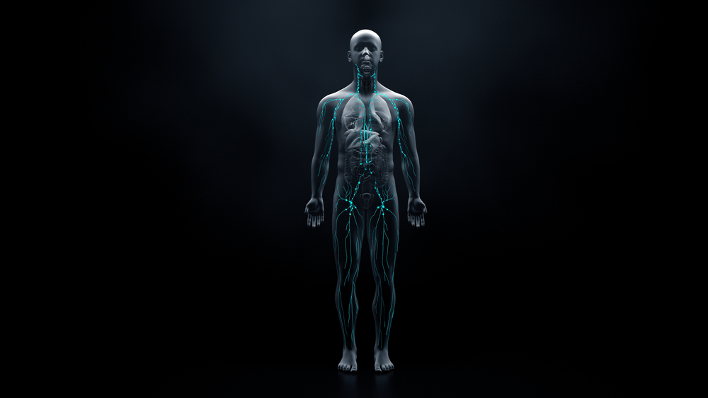
The students’ discovery also raises an important question about how future doctors learn anatomy. Medical schools increasingly use digital resources and synthetic models to help students understand the body’s structures. These tools can make complicated concepts easier to visualize, particularly during the early stages of medical training.
However, standardized models cannot always represent the full range of natural differences found in human bodies. Real donated specimens allow students to observe variations that may not appear in textbooks or educational diagrams. Roy and Bruce encountered the new attachment because they were examining actual human anatomy rather than relying exclusively on an idealized representation.
Tubbs has emphasized that anatomy remains a developing scientific discipline. He also serves as co-editor-in-chief of Gray’s Anatomy, one of the world’s best-known medical reference textbooks. His involvement in the discovery makes the experience particularly interesting, especially because even an experienced anatomist initially struggled to identify the structure.
The finding also gave both students a more personal connection to medical research. Roy explained that the experience strengthened his determination to pursue a career in medicine and encouraged him to continue studying anatomy. Bruce described the project as his first major research experience and continued his academic work in neuroscience at Tulane.
Their experience shows how hands-on learning can encourage students to question established knowledge. A textbook provides an essential foundation, but careful observation and scientific investigation allow future medical professionals to contribute new information.
Human Anatomy Continues to Evolve as a Field of Research
The discovery of the iliac attachment of the psoas major offers an interesting example of how scientific knowledge develops. Researchers had studied the hip region for generations, yet two undergraduate students noticed a structure that had not previously been documented in humans.
The finding does not mean that existing medical knowledge is suddenly incorrect. Instead, it adds another anatomical variation that researchers can investigate and potentially incorporate into future medical education. The next stage will involve determining whether the structure can be identified in living people and whether it has any meaningful relationship with hip health.
For now, there is no reason for people to assume that this newly identified attachment is responsible for their hip discomfort or that they need a different treatment approach. Its clinical significance remains uncertain, and researchers still have substantial work ahead.
What began as an unfamiliar strip of muscle in a university anatomy laboratory has become a published scientific discovery. If future studies establish a connection between this attachment and certain hip conditions, the finding could eventually help doctors make more informed decisions about diagnosis, rehabilitation, and surgery.