A woman in her 50s has received a custom-made, 3D-printed windpipe after surgery to remove thyroid cancer left part of her original airway missing. The procedure, performed in South Korea, marks a significant development in medical technology, combining living cells, specialised printing equipment, and years of scientific research to create a replacement designed specifically for her body.
Even more striking, the artificial windpipe contains cells obtained from other patients, alongside a biodegradable framework intended to support the structure while her body regenerates its own tissue. Six months after the operation, the replacement was reportedly healing well, with new blood vessels beginning to form. The patient also did not require immunosuppressant drugs, raising hopes that this approach could eventually offer new possibilities for people who have lost sections of their windpipe.
How Doctors Created A Replacement Windpipe
The pioneering operation took place at Seoul St Mary’s Hospital in 2023, following a collaboration between the Catholic University of Korea, Gachon University, and biomedical engineering company T&R Biofab. The patient had undergone surgery to remove thyroid cancer, which left her without part of her trachea. This tube carries air from the throat towards the lungs, making its structure essential for breathing. Replacing a missing section presents a difficult medical challenge because the new structure must maintain an open airway while allowing the surrounding tissues to heal and function properly.
To create a replacement that matched the patient’s anatomy, the medical team used CT and MRI scans to determine the required dimensions. The resulting windpipe measured less than 5 centimetres, or approximately 2 inches, in length. Rather than relying on a standard device, the researchers designed a structure specifically for this patient, accounting for the size and shape of the missing section. The printing process took less than two weeks, and the transplant was completed during a surgical procedure lasting approximately half a day.
The procedure brought together decades of work in biomedical engineering and tissue research. Scientists had been investigating the possibilities of this technology since at least 2004, building laboratory evidence before progressing towards human application. The operation represented a major step in applying those efforts to a patient who had lost part of an essential airway. However, further research is needed to establish how reliably the technique works and whether the results can be maintained over many years.
What Is Inside The 3D-Printed Windpipe?
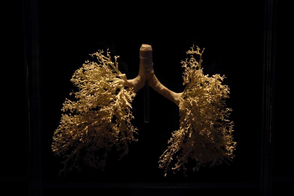
The artificial windpipe combines biological material with a manufactured supporting structure. Researchers used nasal stem cells and cartilage cells obtained from other patients, including tissue discarded during procedures for nasal congestion and nasal septum surgery. These cells were used to create components of the replacement, including cartilage and a mucosal lining. The mucosa is the moist tissue that lines parts of the respiratory system, including the nose and airways, where it helps protect the underlying surfaces.
The structure also contains polycaprolactone, known as PCL, a biodegradable material that provides physical support. Another essential component is bio-ink, which carries living cells during the bioprinting process. Conventional 3D printing typically produces objects from materials such as plastic or metal, whereas bioprinting can incorporate biological components into engineered structures. Combining these materials allowed the researchers to create a customised replacement that incorporated living cells while retaining enough structural support for implantation.
Using cells from other patients was a notable feature of the procedure because the biological material was not collected directly from the woman receiving the transplant. The team also reported that she did not need immunosuppressant drugs after the operation. Such medication is commonly used to reduce the immune system’s response against transplanted tissue. Although avoiding these drugs could offer advantages, one patient’s experience cannot establish that future recipients will have the same outcome or avoid immune-related complications.
Why The Artificial Windpipe Has A Five-Year Lifespan
One of the most important details concerns the biodegradable framework supporting the replacement. The PCL material is expected to last approximately five years before breaking down, according to the reported design. During that period, researchers hope the patient’s body will gradually regenerate its own tracheal tissue, allowing natural tissue to take over some of the structural functions initially provided by the artificial material.
This approach could eventually offer a different way to reconstruct damaged organs. Instead of relying entirely on a permanent artificial replacement, doctors would provide a structure designed to support the body’s own regenerative processes. The concept depends on the patient’s tissue developing sufficiently to maintain the airway as the biodegradable framework breaks down. That outcome remains a goal rather than a guaranteed result, and the long-term performance of the transplant will be crucial in determining whether the approach is successful.

The five-year estimate also refers to the expected lifespan of the PCL component, not a confirmed expiration date for the entire transplanted windpipe. Researchers still need to understand how the replacement behaves over extended periods and whether regenerated tissue can maintain normal airway function. If the approach proves durable, it could become relevant to patients whose tracheas have been damaged by cancer surgery, congenital abnormalities, or serious injuries. For now, the procedure represents an early step towards that possibility.
The Patient’s Recovery Revealed An Encouraging Development
Six months after the transplant, the patient’s replacement windpipe was reportedly healing well, with new blood vessels beginning to form. Blood vessels supply oxygen and nutrients to living tissue, making their development an encouraging observation as the team monitored the transplanted structure. These early findings suggested that biological activity was taking place around the replacement, although longer follow-up is needed to establish whether the tissue can continue developing and maintain the airway successfully.
The absence of a requirement for immunosuppressant drugs was another notable aspect of the case. Such drugs can carry risks because they reduce the immune system’s ability to respond to infections and other threats. Avoiding them could make a similar procedure more attractive if future research confirms that the approach can be performed safely and consistently. However, the experience of a single patient cannot establish the likelihood of complications or predict how other recipients might respond.
The research was still undergoing peer review for potential publication in a scientific journal when the procedure was reported in March 2024. Peer review allows other researchers to assess a study’s methods, evidence, and conclusions. The procedure was described as a world-first achievement, but the evidence available at that stage did not establish that the technique was ready for routine use. The patient’s recovery offered encouraging early observations, while the long-term outcome remained an essential unanswered question.
How Specialised Printers Can Produce Living Structures
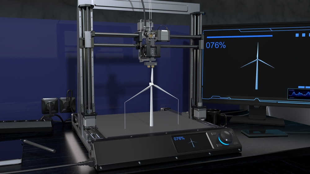
Creating a replacement windpipe requires equipment capable of producing a hollow tube with precise dimensions. The structure must remain open to allow air to pass through, and it must fit the recipient’s anatomy closely enough to function as intended. T&R Biofab developed a specialist printer designed to manufacture hollow tubular structures with high precision, making it suitable for the technical requirements of the procedure.
The machine was developed specifically for Seoul St Mary’s Hospital, meaning it was not a standard printer that any hospital could immediately purchase and use. Expanding the technique would require suitable equipment, trained teams, and a reliable manufacturing process. The technology also depends on carefully handling biological materials and ensuring that the printed structure meets the requirements for implantation. Producing the right shape is only one part of the challenge; the replacement must also remain functional inside a living body.
The development process included laboratory studies and animal testing, including research involving beagles. This work helped build the evidence needed for regulatory assessment before the technology could progress towards a human procedure. The collaboration between medical researchers, engineers, and a specialist manufacturing company demonstrates how complex medical innovations can require expertise from several fields. Wider use will depend on further testing, regulatory decisions, and evidence that the procedure can produce consistent results for different patients.
Could 3D-Printed Organs Change Transplant Medicine?
The shortage of donor organs has encouraged researchers to investigate technologies that might eventually provide alternatives to conventional transplantation. Bioprinting offers one possible route by allowing scientists to construct biological structures using living cells and supporting materials. However, creating a functional replacement for a complex human organ remains far more difficult than printing a medical model or a simple structure. A windpipe must maintain an open passage for air, support living tissue, and integrate with the surrounding anatomy.
Dr Paulo Marinho, head of scientific strategy at T&R Biofab, described the potential of the technology while acknowledging that its limitations remain significant. “While it’s too soon to say that 3D-bioprinting could be the solution for the current shortage of organs for transplantation, it definitely increases the hopes to partially solve the issue for some organs or some specific indications, or at least fill the gap between classic medical devices and organ transplants,” Marinho said.
His assessment reflects the cautious optimism surrounding bioprinting. Rather than replacing conventional transplantation immediately, the technology could eventually provide alternatives for specific conditions or patients with limited treatment options. Researchers have also investigated printed heart patches made from stem-cell-derived material, with studies examining their potential to assist damaged hearts in rats. Such work represents a different application of bioprinting, since supporting injured tissue is not the same as replacing an entire functioning organ.
What Researchers Must Prove Before Wider Use
The Korean windpipe transplant demonstrated that a customised structure incorporating living cells could be manufactured and implanted into a human patient. However, several questions remain before the technique can become a widely available treatment. Researchers need to determine how long the replacement remains functional, whether regenerated tissue can maintain the airway as the supporting material degrades, and how patients fare over extended periods. They must also establish whether the approach can work consistently across people with different medical histories and anatomical requirements.
Access presents another challenge because the procedure relied on specialist equipment developed for one hospital. Wider adoption would require a more scalable manufacturing process, appropriate clinical facilities, and evidence that the benefits outweigh the risks. Regulatory approval and further clinical research will also be necessary before doctors can determine which patients might benefit most. The reported procedure offers a potential direction for future treatment, but it does not yet establish a routine solution for people who have lost part of their windpipe.
For patients facing complex airway reconstruction, the possibility of a replacement designed around their own anatomy could eventually expand the available treatment options. The next major milestone will be demonstrating that the transplanted structure can remain functional over time while the body develops enough natural tissue to support breathing. That evidence will determine whether this pioneering operation becomes the foundation for a broader medical technique or remains an exceptional achievement in early-stage research.
Sources:
- Pubmed.ncbi.nlm.nih.gov. (n.d.). https://pubmed.ncbi.nlm.nih.gov/38646888/?
- Park, J., Yoon, J., Lee, J. B., Shin, Y. M., Lee, K., Bae, S., Lee, J., Yu, J., Jung, C., Youn, Y., Kim, H., & Kim, D. (2019). Experimental Tracheal Replacement Using 3-dimensional Bioprinted Artificial Trachea with Autologous Epithelial Cells and Chondrocytes. Scientific Reports, 9(1), 2103. https://doi.org/10.1038/s41598-019-38565-z