A baby facing a potentially dangerous lung tumor received treatment before birth, giving doctors a chance to reduce the pressure on the developing heart and lungs. The procedure used a thin needle and microwave energy while the baby was still inside the womb.
The treatment was performed at 22 weeks of pregnancy after scans showed a rapidly growing lung abnormality measuring about 5 centimeters. Within three days, the tumor had shrunk significantly and the dangerous fluid buildup in the baby’s abdomen had disappeared.
Doctors Faced A Serious Problem At 22 Weeks
The case involved a 40-year-old woman whose baby was diagnosed with microcystic congenital pulmonary airway malformation, commonly known as CPAM, during the 22nd week of pregnancy. CPAM is a rare congenital condition involving abnormal development of lung tissue during pregnancy, and most cases do not become life-threatening.
Severe cases can be very different, however, particularly when the abnormal tissue grows rapidly. A large lesion can take up much of the space inside the fetal chest, placing pressure on the developing lungs and heart and potentially causing serious complications before birth.
In this pregnancy, ultrasound scans showed that the abnormal growth had reached approximately 5 centimeters and occupied much of the baby’s right chest cavity. The mass was compressing the heart and lungs, and doctors also detected ascites, which is a buildup of fluid inside the abdomen.
The combination made the situation particularly concerning because the pregnancy was still relatively early. The baby needed additional time to develop, but the growing lesion was already interfering with important organs.
According to the supplied reports, CPAM occurs in roughly one in 25,000 to 35,000 pregnancies. While the condition is usually manageable, rapidly enlarging lesions can create severe problems in a small number of pregnancies.
When A Lung Lesion Becomes Dangerous
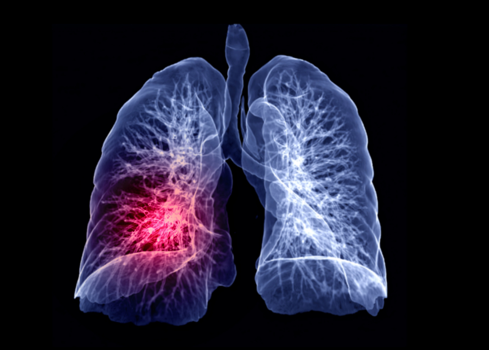
The main concern with a large CPAM is the amount of space the abnormal tissue occupies inside the fetal chest. A rapidly growing lesion can compress structures that the baby needs for normal development, including the heart and healthy portions of the lungs.
Severe cases can also cause abnormal fluid accumulation. The buildup may occur in the abdomen, as it did in this case, or become more widespread in a complication known as hydrops.
That is why doctors closely monitor pregnancies involving significant CPAM. The size of the lesion, how quickly it grows, its internal structure, and its effect on the fetus can all influence what happens next.
Some babies with CPAM require little or no intervention before birth, while others need specialized fetal treatment. The difference can depend heavily on how the lesion behaves during pregnancy.
Why Conventional Treatment Was Not An Option
Doctors already have several ways to manage severe fetal lung lesions, but those approaches are not appropriate for every type of CPAM. Treatment has to match the physical characteristics of the abnormal tissue and the risks facing the mother and baby.
Some CPAMs contain large, fluid-filled cysts that can be drained using a catheter. In certain situations, doctors can place a drainage tube through the mother’s abdomen and uterus so that fluid can leave the cyst and reduce pressure inside the fetal chest.
Another approach involves targeting the blood supply to the abnormal tissue. If a lesion has a major feeding vessel, doctors may be able to interrupt that blood supply and reduce the growth.
The lesion in this case presented a different challenge because it consisted mainly of tiny cysts and did not have a central blood vessel that doctors could target. The standard approaches therefore did not offer an obvious solution.
There is also a much more invasive option known as fetal lobectomy. The procedure involves surgically opening the uterus to remove the affected tissue before the fetus is returned to the womb, making it a major fetal operation with significant risks.
The supplied reports cite previous research indicating that survival following this type of intervention has been around 50% in the specific context discussed. That helped underline why the medical team wanted to consider a less invasive alternative for this pregnancy.
How Microwave Ablation Reached The Tumor

The medical team from Shenzhen Baoan Women’s and Children’s Hospital worked with specialists from the Chinese University of Hong Kong to perform the procedure. Their approach involved microwave ablation, a technique that uses controlled heat to destroy targeted abnormal tissue.
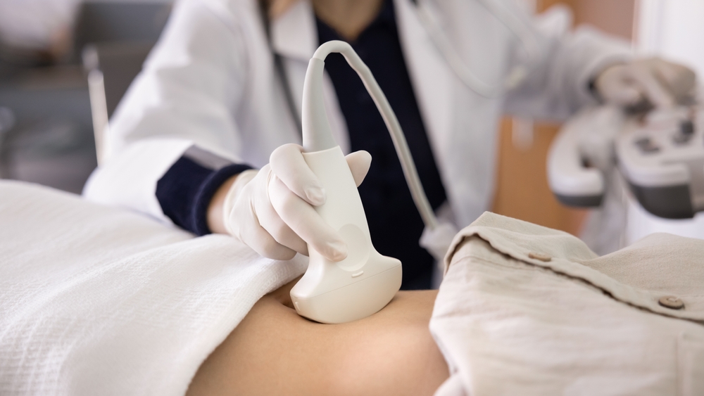
Doctors first used high-definition, real-time ultrasound imaging to identify the tumor and plan the safest route for the needle. The imaging allowed them to see the fetus, the tumor, and surrounding structures throughout the procedure.
A thin ablation needle was then carefully guided through the mother’s abdomen and uterine wall before entering the baby’s chest. The needle was positioned directly inside the abnormal lung tissue so that microwave energy could be delivered to the target.
The approach avoided the need to open the uterus through conventional surgery. Instead, the medical team could reach the tumor through a narrow needle while continuously watching the procedure with ultrasound.
How The Heat Destroyed Abnormal Tissue
Microwave ablation works by using electromagnetic energy to generate heat inside tissue. The energy causes water molecules within the tissue to move rapidly, producing temperatures high enough to damage and destroy the targeted cells.
In this procedure, the active heating stage lasted approximately two minutes, according to the supplied reports. Doctors carefully monitored the heat distribution because the goal was to destroy the abnormal tissue while protecting the baby’s developing organs.
That level of image guidance was important because the tumor was located inside the fetal chest, close to structures that could not simply be moved out of the way. The medical team needed to deliver enough energy to affect the lesion without causing unnecessary damage to healthy tissue.
The procedure therefore combined a very small physical access point with continuous imaging and targeted energy delivery. The result was a treatment designed specifically for a lesion that could not be addressed effectively through the more established options.
The Baby Improved Within Three Days

The first signs of improvement appeared quickly after the procedure. Three days later, scans showed that the tumor had decreased significantly in size, while the fluid buildup in the baby’s abdomen had completely disappeared.
That change reduced the immediate pressure created by the lesion and allowed the pregnancy to continue under medical monitoring. Instead of requiring immediate delivery or more invasive fetal surgery, the medical team could continue watching the pregnancy as the baby developed.
The pregnancy eventually reached 37 weeks. The baby was delivered by Caesarean section and weighed approximately 2.9 kilograms, or about 6.4 pounds.
The newborn received an Apgar score of 10. The Apgar assessment is a quick evaluation performed after birth to assess a newborn’s physical condition, including factors such as breathing, heart rate, muscle tone, reflex response, and skin color.
According to the supplied reports, the infant remained symptom-free at four months of age. The baby had also not required further surgical intervention during that period.
For a pregnancy that had involved a rapidly growing lesion and fluid accumulation at 22 weeks, reaching 37 weeks represented a substantial change in the clinical picture.
What Makes This Procedure Different

The most notable feature of the treatment is that doctors were able to target the fetal tumor without performing conventional open fetal surgery. A narrow needle provided access to the lesion, while ultrasound gave the team continuous visual guidance.
The technique also offered several characteristics that could make it useful for carefully selected cases if future research confirms its safety and effectiveness.
- It uses a minimally invasive approach: Rather than opening the uterus surgically, doctors accessed the tumor through a thin needle inserted through the mother’s abdomen and uterine wall.
- It targets the abnormal tissue directly: Microwave energy was delivered inside the tumor rather than affecting a larger surrounding area.
- It relies on real-time imaging: Ultrasound allowed doctors to monitor the needle position and observe the procedure while it was taking place.
- It produced a rapid response: The supplied reports state that the tumor had shrunk significantly within three days of treatment.
- It allowed the pregnancy to continue: After the procedure, the baby remained in the womb until delivery at 37 weeks.
These features could make microwave ablation an interesting area for further research in fetal medicine. However, the successful outcome of one pregnancy cannot establish that the technique will produce the same results in other babies.
The Procedure Is Promising, But More Evidence Is Needed

The doctors involved described the procedure as a “new milestone” in fetal medicine, according to the supplied reports. The case was also reported in the peer-reviewed journal Ultrasound in Obstetrics & Gynecology on August 20.
That publication provides an opportunity for other specialists to examine the details of the treatment and consider how the approach might be evaluated in future cases. The significance of the report comes from the unusual combination of the fetal condition, the limitations of standard treatments, and the successful use of microwave ablation.
Still, the medical team emphasized an important limitation. The safety and effectiveness of the procedure cannot be conclusively established from a single case.
That caution matters because fetal procedures carry risks that may not become apparent in one successful pregnancy. Doctors need evidence from additional cases to determine whether the technique can be performed consistently and whether complications could emerge when it is used more widely.
Researchers will also need to establish which CPAM lesions are suitable for microwave ablation. The size and structure of the lesion, its location, its relationship with nearby organs, and the condition of the fetus could all influence whether the procedure is appropriate.
A Successful Case Is Not Yet A Standard Treatment
For now, microwave ablation should be viewed as a possible treatment approach under investigation rather than an established solution for severe CPAM.
The distinction is particularly important for parents who may encounter headlines describing the procedure as a breakthrough. A successful individual case can show what may be possible, but it cannot tell doctors how often the approach will work or what risks might appear in a larger group of patients.
Future research will need to examine both benefits and complications. Specialists will also need to determine whether the procedure can be reproduced safely at other medical centers with appropriate fetal medicine expertise.
If those studies produce consistent results, microwave ablation could eventually become another option for a carefully selected group of pregnancies.
What Parents Should Know About CPAM

A diagnosis of CPAM does not automatically mean that a baby will face a severe medical outcome. The condition covers a broad range of abnormalities, and many cases are manageable with monitoring and appropriate care.
The more serious concern arises when a lesion grows quickly and begins affecting nearby organs. A large mass inside the fetal chest can interfere with the development of the lungs and place pressure on the heart, while associated fluid buildup can create additional complications.
That is why prenatal monitoring is so important after a CPAM diagnosis. Doctors can use imaging to follow the size and structure of the lesion and determine whether it is changing the baby’s condition.
Parents may also encounter different treatment recommendations depending on what the scans show. A small or stable lesion may be monitored, while a rapidly growing lesion causing significant compression may require intervention from a specialized fetal medicine team.
The treatment described in this case was considered because conventional options were unsuitable for the specific structure of the tumor. It should therefore not be interpreted as a procedure that every baby with CPAM would need or benefit from.
Treatment Decisions Depend On The Individual Pregnancy
Fetal medicine is highly individualized because doctors must consider both maternal and fetal health at every stage. The same diagnosis can present very differently from one pregnancy to another.
Factors such as gestational age, lesion size, growth rate, location, fluid accumulation, and the effect on the heart and lungs can all influence clinical decisions.
For families receiving a prenatal diagnosis, the most useful step is understanding what the imaging shows and what the medical team believes it means for that particular pregnancy. Treatment decisions should be based on the baby’s individual circumstances rather than on a single case reported in the news.
A New Direction For Fetal Medicine
The Chinese team’s experience provides an example of how doctors are exploring less invasive ways to treat serious conditions before birth. In this case, a thin needle and carefully controlled microwave energy were used to reduce a lung lesion that was already threatening the baby’s developing organs.
The pregnancy continued for another 15 weeks after the procedure, and the baby was delivered at 37 weeks with an Apgar score of 10. The infant was reported to remain symptom-free at four months without requiring additional surgery.
Those results are encouraging, but the next step is evidence. If further research confirms that microwave ablation can be performed safely and consistently, the technique could give fetal medicine specialists another option for some of the most difficult CPAM cases.
For now, the case offers something more useful than a promise of a universal treatment: evidence that carefully targeted intervention before birth may give some babies a better chance to keep developing until delivery.